Copyright
©The Author(s) 2017.
World J Gastroenterol. Sep 7, 2017; 23(33): 6181-6186
Published online Sep 7, 2017. doi: 10.3748/wjg.v23.i33.6181
Published online Sep 7, 2017. doi: 10.3748/wjg.v23.i33.6181
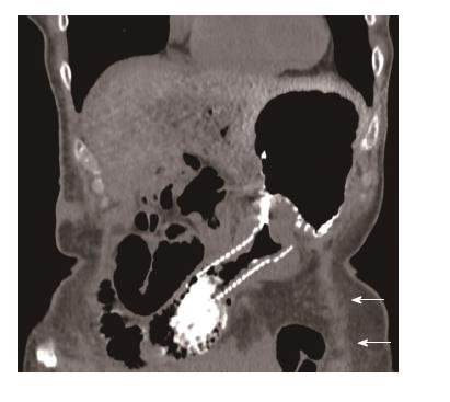
Figure 1 Coronal computed tomography image of a non-covered metallic stent with peroral contrast in the dilated afferent limb in a patient with classical Whipple’s duodenopancreatectomy.
Note the obstructed empty alimentary limb underneath the stomach (arrows).
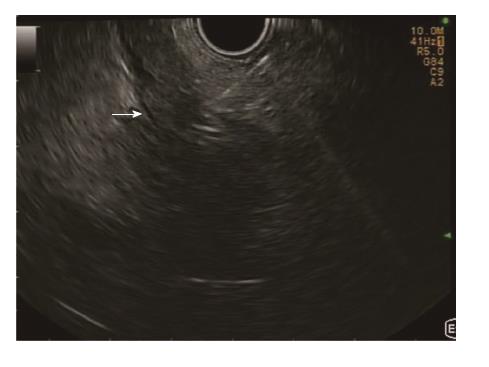
Figure 2 Linear endoscopic ultrasound image of the transgastric puncture of the obstructed alimentary limb (arrow) with a 19 gauche needle.
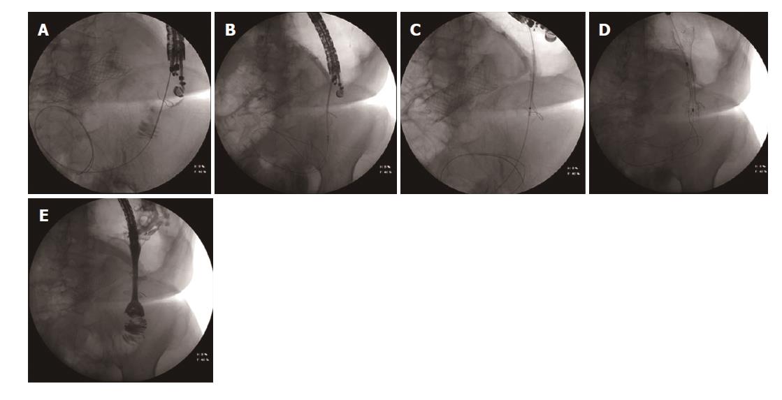
Figure 3 Fluoroscopic image.
A: The transgastric access of the alimentary limb using a 19 gauche needle. Correct access of the limb was confirmed after injection of contrast dye. A: 0.035 inch guidewire is introduced into the alimentary limb. Note the SEMS in the afferent limb; B: The HotAxios lumen apposing stent deployment between the alimentary limb and the gastric lumen; C: The HotAxios lumen apposing stent migrating outside to stomach. The distal flange is well positioned inside the lumen of the alimentary limb; D: The fully covered oesophageal stent inside the HotAxios lumen apposing stent, creating a gastrojejunostomy; E: Fluoroscopic image confirming the creation of a gastrojejunostomy between the stomach and the alimentary limb using the combination of a HotAxios lumen opposing stent and a fully covered oesophageal stent. Water soluble contrast dye is injected through the oesophageal stent into the alimentary limb. Note the non-covered SEMS in the afferent limb.
Figure 4 Fluoroscopic image.
A: A percutaneous transhepatic cholangiography confirming patency of the biliary anastomosis. The malignant stricture (arrow) is located at the level of the afferent limb (afferent limb syndrome), leading to dilated intrahepatic bile ducts and a dilatation of the afferent limb. Note the presence of an old Ovesco clip in the stomach. B: The single-balloon enteroscopy up to the level of the malignant stricture in the afferent limb. Contrast injection through the working channel of the enteroscope clearly delineates the length and the malignant aspect of the stricture (arrow). C: The peroral insertion technique of the self-expandable metal stent over a stiff guidewire through the overtube with the balloon inflated to protect the intestinal mucosa and to guide the catheter up to the malignant stricture of the afferent limb. D: The self-expandable metal stent in place in the malignant stricture of the afferent limb. Both the stiff guidewire and the overtube of the single-balloon enteroscope were used to insert the SEMS. SEMS: Self-expandable metal stent.
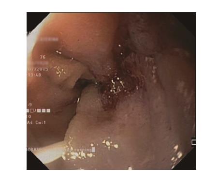
Figure 5 Endoscopic image of the malignant stricture of the afferent limb.
Note the guidewire inserted into the stricture.
- Citation: Mouradides C, Taha A, Borbath I, Deprez PH, Moreels TG. How to treat intestinal obstruction due to malignant recurrence after Whipple’s resection for pancreatic head cancer: Description of 2 new endoscopic techniques. World J Gastroenterol 2017; 23(33): 6181-6186
- URL: https://www.wjgnet.com/1007-9327/full/v23/i33/6181.htm
- DOI: https://dx.doi.org/10.3748/wjg.v23.i33.6181


