Copyright
©The Author(s) 2017.
World J Gastroenterol. Sep 7, 2017; 23(33): 6181-6186
Published online Sep 7, 2017. doi: 10.3748/wjg.v23.i33.6181
Published online Sep 7, 2017. doi: 10.3748/wjg.v23.i33.6181
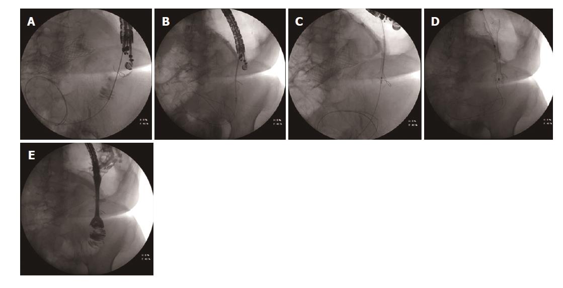
Figure 3 Fluoroscopic image.
A: The transgastric access of the alimentary limb using a 19 gauche needle. Correct access of the limb was confirmed after injection of contrast dye. A: 0.035 inch guidewire is introduced into the alimentary limb. Note the SEMS in the afferent limb; B: The HotAxios lumen apposing stent deployment between the alimentary limb and the gastric lumen; C: The HotAxios lumen apposing stent migrating outside to stomach. The distal flange is well positioned inside the lumen of the alimentary limb; D: The fully covered oesophageal stent inside the HotAxios lumen apposing stent, creating a gastrojejunostomy; E: Fluoroscopic image confirming the creation of a gastrojejunostomy between the stomach and the alimentary limb using the combination of a HotAxios lumen opposing stent and a fully covered oesophageal stent. Water soluble contrast dye is injected through the oesophageal stent into the alimentary limb. Note the non-covered SEMS in the afferent limb.
- Citation: Mouradides C, Taha A, Borbath I, Deprez PH, Moreels TG. How to treat intestinal obstruction due to malignant recurrence after Whipple’s resection for pancreatic head cancer: Description of 2 new endoscopic techniques. World J Gastroenterol 2017; 23(33): 6181-6186
- URL: https://www.wjgnet.com/1007-9327/full/v23/i33/6181.htm
- DOI: https://dx.doi.org/10.3748/wjg.v23.i33.6181


