Copyright
©The Author(s) 2017.
World J Gastroenterol. Aug 21, 2017; 23(31): 5755-5763
Published online Aug 21, 2017. doi: 10.3748/wjg.v23.i31.5755
Published online Aug 21, 2017. doi: 10.3748/wjg.v23.i31.5755
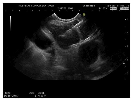
Figure 1 Endoscopic ultrasound-guided fine-needle biopsy of an intra-abdominal lymph node.
The needle and needle tip are clearly visible inside the targeted lymph node.
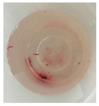
Figure 2 Example of a core sample obtained with endoscopic ultrasound-guided tissue acquisition using a biopsy needle.
Figure 3 Small cell carcinoma from an fine-needle aspiration subcarinal lymph node (cell block).
A: Note the small cell neoplastic population with hyperchromatic nuclei, scant cytoplasm and absent nucleoli; B: Nuclear positivity for TTF-1; C: Cytoplasmic positivity for synaptophysin.
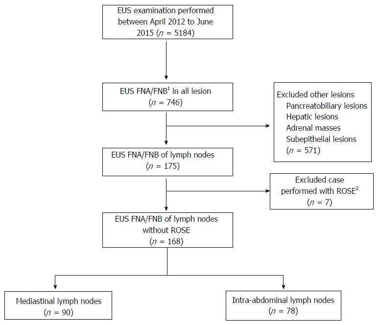
Figure 4 Flow chart of the selection of endoscopic ultrasound-guided tissue acquisition cases.
1Fine-needle aspiration/biopsy; 2Rapid on-site evaluation. EUS: endoscopic ultrasound; FNA: Fine-needle aspiration; FNB: Fine-needle biopsy.
- Citation: Chin YK, Iglesias-Garcia J, de la Iglesia D, Lariño-Noia J, Abdulkader-Nallib I, Lázare H, Rebolledo Olmedo S, Dominguez-Muñoz JE. Accuracy of endoscopic ultrasound-guided tissue acquisition in the evaluation of lymph nodes enlargement in the absence of on-site pathologist. World J Gastroenterol 2017; 23(31): 5755-5763
- URL: https://www.wjgnet.com/1007-9327/full/v23/i31/5755.htm
- DOI: https://dx.doi.org/10.3748/wjg.v23.i31.5755


