Copyright
©The Author(s) 2017.
World J Gastroenterol. Mar 14, 2017; 23(10): 1843-1850
Published online Mar 14, 2017. doi: 10.3748/wjg.v23.i10.1843
Published online Mar 14, 2017. doi: 10.3748/wjg.v23.i10.1843
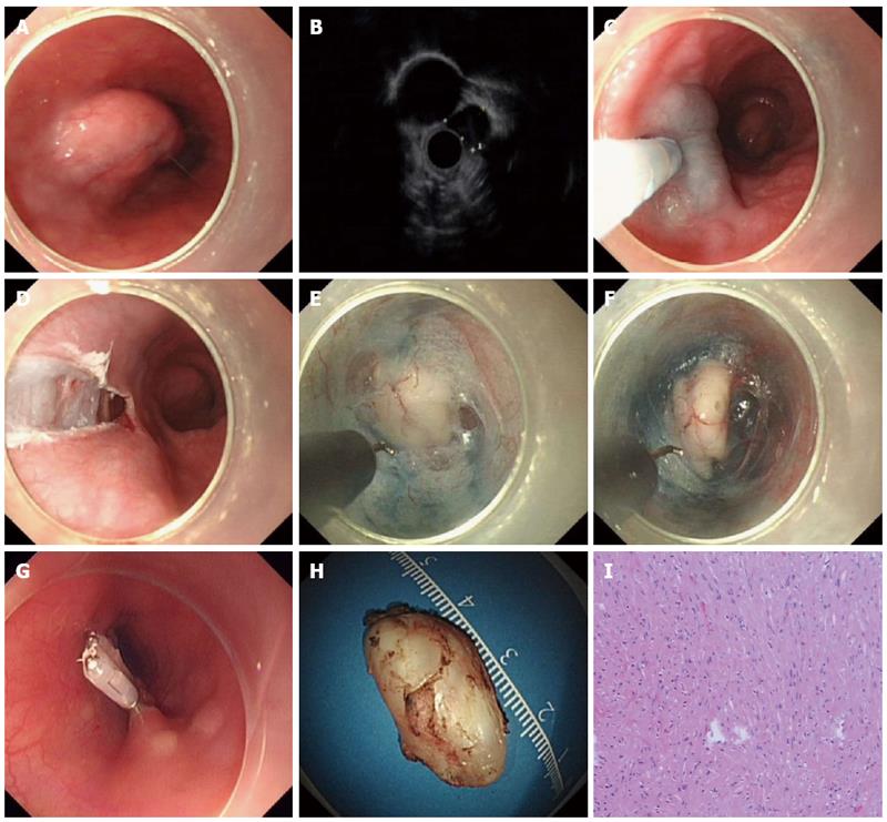
Figure 1 Illustration of endoscopic submucosal tunnel dissection for esophageal submucosal tumor in a patient using a hook knife.
A: An submucosal tumor (SMT) located at the left lateral wall of the mid esophagus; B: Endoscopic ultrasonography revealed a 32.8 mm × 14.7 mm hypoechoic submucosal lesion; C and D: A 2-cm longitudinal incision is made into the mucosa after injection of natural saline with indigo carmine and epinephrine; E: After a mucosal incision was made, submucosal dissection was made approximately 4 cm proximal to the SMT with a hook knife, creating a submucosal tunnel until the tumor was visible; F: Dissection was done along the margin of the tumor; G: Endoclips were used to close the entry of the submucosal tunnel; H and I: Pathological examination revealed that the resected specimen was a 28-mm leiomyoma.
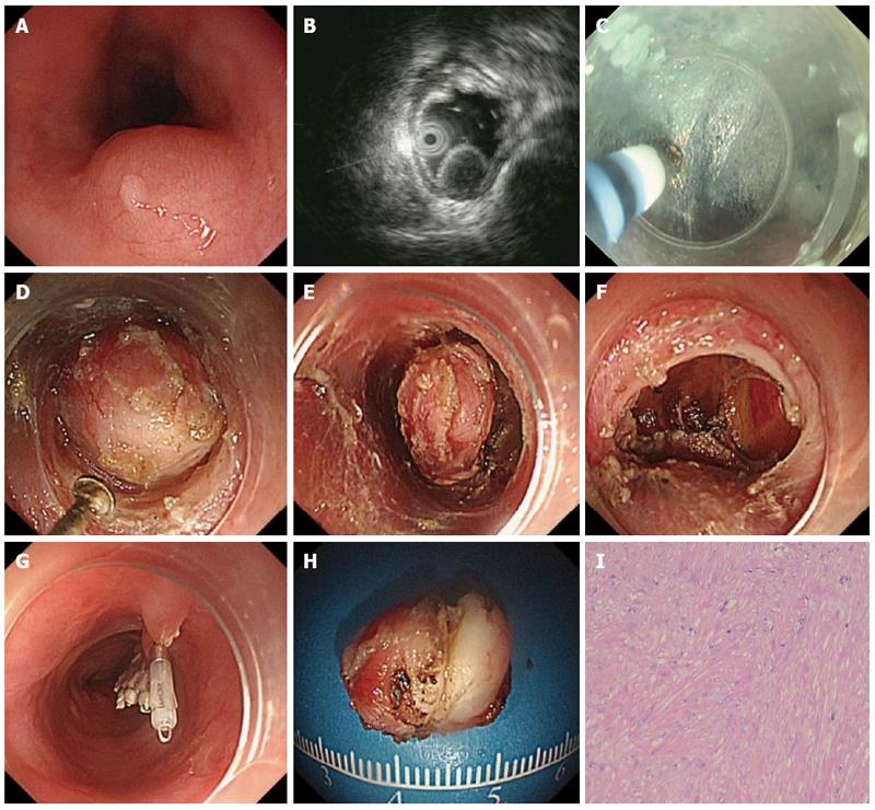
Figure 2 Illustration of endoscopic submucosal tunnel dissection for esophageal submucosal tumor in a patient using a hybrid knife.
A: An submucosal tumor (SMT) located at the posterior wall of the mid esophagus; B: Endoscopic ultrasonography revealed a 15.2 mm × 11.4 mm hypoechoic submucosal lesion; C: After a 2-cm longitudinal mucosal incision was made, submucosal dissection was made approximately 4 cm proximal to the SMT with a hybrid knife, creating a submucosal tunnel; D and E: Dissection was done along the margin of the tumor; F: Endoscopic view of submucosal tunnel after removal of the tumor; G: Endoclips were used to close the entry of the submucosal tunnel; H and I: Pathological examination revealed that the resected specimen was a 15-mm leiomyoma.
- Citation: Zhou JQ, Tang XW, Ren YT, Wei ZJ, Huang SL, Gao QP, Zhang XF, Yang JF, Gong W, Jiang B. Endoscopic submucosal tunnel dissection of upper gastrointestinal submucosal tumors: A comparative study of hook knife vs hybrid knife. World J Gastroenterol 2017; 23(10): 1843-1850
- URL: https://www.wjgnet.com/1007-9327/full/v23/i10/1843.htm
- DOI: https://dx.doi.org/10.3748/wjg.v23.i10.1843


