Copyright
©The Author(s) 2016.
World J Gastroenterol. Mar 7, 2016; 22(9): 2678-2700
Published online Mar 7, 2016. doi: 10.3748/wjg.v22.i9.2678
Published online Mar 7, 2016. doi: 10.3748/wjg.v22.i9.2678
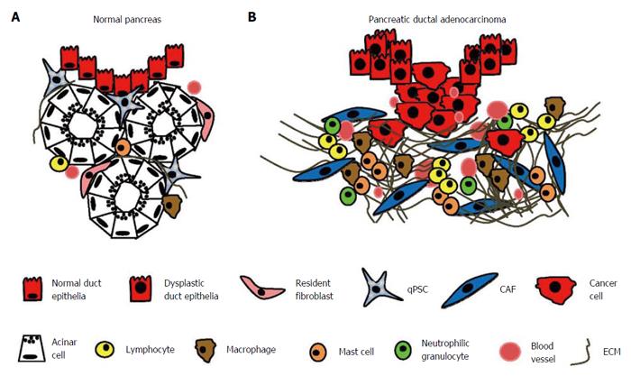
Figure 1 Desmoplastic reaction in pancreatic ductal adenocarcinoma.
The normal exocrine pancreas consists of acini with acinar cells and pancreatic ducts lined by epithelial cells. Quiescent pancreatic stellate cells (qPSCs) and interlobular fibroblasts are located in the periacinar space. Only a few T cells are observed, and B lymphocytes, plasma cells, and eosinophilic and neutrophilic granulocytes are very rare. The extracellular matrix (ECM) is largely limited to thin interlobular septa and pancreatic ducts (A); In pancreatic ductal adenocarcinoma, cancer cells permeate the basal membrane of dysplastic pancreatic ducts and invade the surrounding tissue. This invasion is accompanied by a strong desmoplastic reaction in which cancer-associated fibroblasts (CAFs), arising mainly from quiescent PSCs, synthesize an abundance of ECM proteins. Lymphocytes, macrophages, and mast cells infiltrate the peritumoral stroma. There is an increased need for oxygen and nutrients, leading to increased angiogenesis (B).
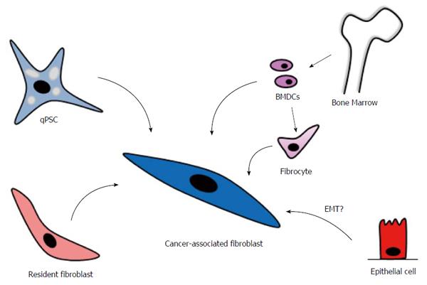
Figure 2 Cellular sources to cancer-associated fibroblasts in pancreatic ductal adenocarcinoma.
Pancreatic stellate cells (PSCs) constitute the most important cellular source of cancer-associated fibroblasts (CAFs) in pancreatic cancer. In the normal pancreas, quiescent PSCs (qPSCs) have a periacinar location. When activated by cytokines or oxidative stress, they develop a myofibroblast-like phenotype. Resident periductal and interlobular fibroblasts can also contribute to the CAF population. Moreover, several studies have indicated that bone marrow-derived cells (BMDCs) are recruited to the pancreas during tissue injury, where they gain CAF-like properties. It could also be speculated that epithelial cells, through epithelial-mesenchymal transition (EMT), could be a source of CAFs.
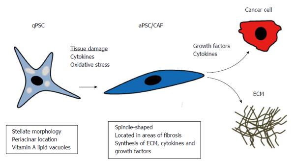
Figure 3 Activation of quiescent pancreatic stellate cells in pancreatic cancer.
In the normal pancreas, pancreatic stellate cells are in the quiescent state (qPSCs) and located in the periacinar space. They have a stellate morphology and contain vitamin A lipid vacuoles. During carcinogenesis, they become activated (aPSCs). aPSCs are the most important cellular source of cancer-associated fibroblasts (CAFs) in PC. CAFs, characterized by a spindle-shaped morphology, are the main effector cells in the desmoplastic reaction, and they synthesize extracellular matrix (ECM) proteins. Further, they produce the growth factors and cytokines that promote cancer cell proliferation and migration.
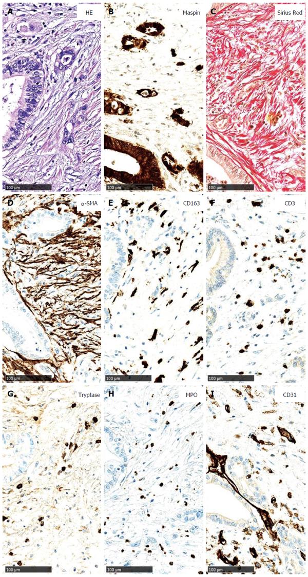
Figure 4 Main components of the tumour stroma in pancreatic cancer.
A: Pancreatic cancer (PC) cells, arranged in small groups and duct-like structures, are surrounded by desmoplastic stroma (HE staining); B: PC cells strongly express maspin (maspin immunostaining); C: Using a Sirius red stain, the collagen fibres of the desmoplastic stroma are highlighted; D: Numerous α-smooth muscle actin-positive cancer-associated fibroblasts are observed; E: Tumour-infiltrating macrophages (CD163 immunostaining); F: T cells (CD3 immunostaining) are shown; G: Additionally, a few mast cells (tryptase immunostaining); H: neutrophilic granulocytes (myeloperoxidase immunostaining) are present; I: Several newly formed small blood vessels are located in the desmoplastic stroma (CD31 immunostaining).
- Citation: Nielsen MFB, Mortensen MB, Detlefsen S. Key players in pancreatic cancer-stroma interaction: Cancer-associated fibroblasts, endothelial and inflammatory cells. World J Gastroenterol 2016; 22(9): 2678-2700
- URL: https://www.wjgnet.com/1007-9327/full/v22/i9/2678.htm
- DOI: https://dx.doi.org/10.3748/wjg.v22.i9.2678


