Copyright
©The Author(s) 2016.
World J Gastroenterol. Mar 7, 2016; 22(9): 2678-2700
Published online Mar 7, 2016. doi: 10.3748/wjg.v22.i9.2678
Published online Mar 7, 2016. doi: 10.3748/wjg.v22.i9.2678
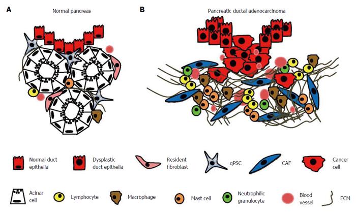
Figure 1 Desmoplastic reaction in pancreatic ductal adenocarcinoma.
The normal exocrine pancreas consists of acini with acinar cells and pancreatic ducts lined by epithelial cells. Quiescent pancreatic stellate cells (qPSCs) and interlobular fibroblasts are located in the periacinar space. Only a few T cells are observed, and B lymphocytes, plasma cells, and eosinophilic and neutrophilic granulocytes are very rare. The extracellular matrix (ECM) is largely limited to thin interlobular septa and pancreatic ducts (A); In pancreatic ductal adenocarcinoma, cancer cells permeate the basal membrane of dysplastic pancreatic ducts and invade the surrounding tissue. This invasion is accompanied by a strong desmoplastic reaction in which cancer-associated fibroblasts (CAFs), arising mainly from quiescent PSCs, synthesize an abundance of ECM proteins. Lymphocytes, macrophages, and mast cells infiltrate the peritumoral stroma. There is an increased need for oxygen and nutrients, leading to increased angiogenesis (B).
- Citation: Nielsen MFB, Mortensen MB, Detlefsen S. Key players in pancreatic cancer-stroma interaction: Cancer-associated fibroblasts, endothelial and inflammatory cells. World J Gastroenterol 2016; 22(9): 2678-2700
- URL: https://www.wjgnet.com/1007-9327/full/v22/i9/2678.htm
- DOI: https://dx.doi.org/10.3748/wjg.v22.i9.2678


