Copyright
©The Author(s) 2016.
World J Gastroenterol. Nov 28, 2016; 22(44): 9803-9812
Published online Nov 28, 2016. doi: 10.3748/wjg.v22.i44.9803
Published online Nov 28, 2016. doi: 10.3748/wjg.v22.i44.9803
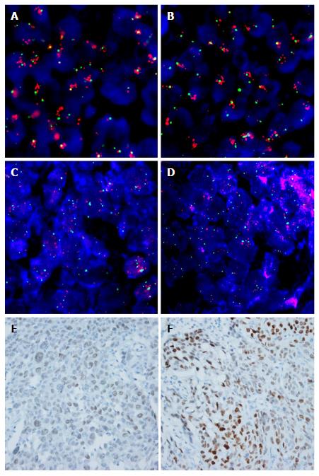
Figure 1 Representative images of fluorescence in situ hybridization and immunohistochemistry for fibroblast growth factor receptor 1 and MYC in esophageal squamous cell carcinoma.
A and B: High amplification of FGFR1 with increased gene copies of FGFR1 (red signal), compared to chromosome 8 (CEP8, green signal), was observed; C and D: High amplification of MYC with increased gene copies of MYC (orange signal), compared to CEP8 was observed; E and F: MYC immunohistochemistry in the nuclei of tumor cells; E: Weak, F: Strong intensity (original magnification, × 400). FGFR1: Fibroblast growth factor receptor 1.
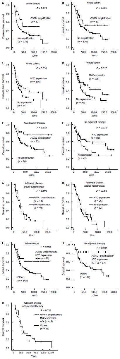
Figure 2 Kaplan-Meier plots and log rank test results.
Disease-free survival (DFS) and overall survival (OS) in patients with resected esophageal squamous cell carcinoma (ESCC); A and B: According to FGFR1 amplification; C and D: According to MYC expression status; OS was also plotted according to FGFR1 amplification and MYC expression statuses; E and F: In patients with ESCC who did not receive adjuvant therapy; G and H: in those with ESCC who received adjuvant therapy; I-K: OS of patients with ESCC according to combined FGFR1 amplification and MYC expression status was plotted and analyzed in all patients with resected ESCC, as well as in those with and without adjuvant therapy, respectively. FGFR1: Fibroblast growth factor receptor 1.
- Citation: Kwon D, Yun JY, Keam B, Kim YT, Jeon YK. Prognostic implications of FGFR1 and MYC status in esophageal squamous cell carcinoma. World J Gastroenterol 2016; 22(44): 9803-9812
- URL: https://www.wjgnet.com/1007-9327/full/v22/i44/9803.htm
- DOI: https://dx.doi.org/10.3748/wjg.v22.i44.9803


