Copyright
©The Author(s) 2016.
World J Gastroenterol. Nov 28, 2016; 22(44): 9803-9812
Published online Nov 28, 2016. doi: 10.3748/wjg.v22.i44.9803
Published online Nov 28, 2016. doi: 10.3748/wjg.v22.i44.9803
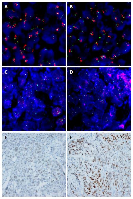
Figure 1 Representative images of fluorescence in situ hybridization and immunohistochemistry for fibroblast growth factor receptor 1 and MYC in esophageal squamous cell carcinoma.
A and B: High amplification of FGFR1 with increased gene copies of FGFR1 (red signal), compared to chromosome 8 (CEP8, green signal), was observed; C and D: High amplification of MYC with increased gene copies of MYC (orange signal), compared to CEP8 was observed; E and F: MYC immunohistochemistry in the nuclei of tumor cells; E: Weak, F: Strong intensity (original magnification, × 400). FGFR1: Fibroblast growth factor receptor 1.
- Citation: Kwon D, Yun JY, Keam B, Kim YT, Jeon YK. Prognostic implications of FGFR1 and MYC status in esophageal squamous cell carcinoma. World J Gastroenterol 2016; 22(44): 9803-9812
- URL: https://www.wjgnet.com/1007-9327/full/v22/i44/9803.htm
- DOI: https://dx.doi.org/10.3748/wjg.v22.i44.9803


