Copyright
©The Author(s) 2016.
World J Gastroenterol. Sep 21, 2016; 22(35): 7926-7937
Published online Sep 21, 2016. doi: 10.3748/wjg.v22.i35.7926
Published online Sep 21, 2016. doi: 10.3748/wjg.v22.i35.7926
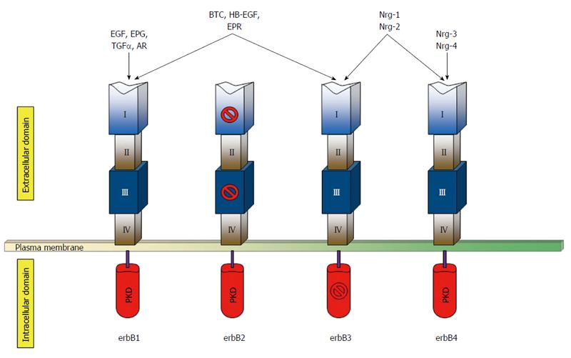
Figure 1 Schematic representation of the human erbB receptors in basal condition.
The extracellular portion of each receptor consists of four domains (I-IV). Both domains I and III, which are related leucine-rich segments, actively participate in ligand binding, except for those of erbB2. Domains II and IV contain numerous cysteine residues and participates in dimer formation. The kinase domain of erbB3 is kinase-impaired. The growth factor groups that bind each receptor are indicated on the top. PKD: Protein kinase domain; EGF: Epidermal growth factor; EPG: Epigen; TGFα: Transforming growth factor-α; AR: Amphiregulin; BTC: Betacellulin; HB-EGF: Heparin-binding epidermal growth-factor like growth factor; EPR: Epiregulin; Nrg-1/2/3/4: Neuregulin-1/2/3/4.
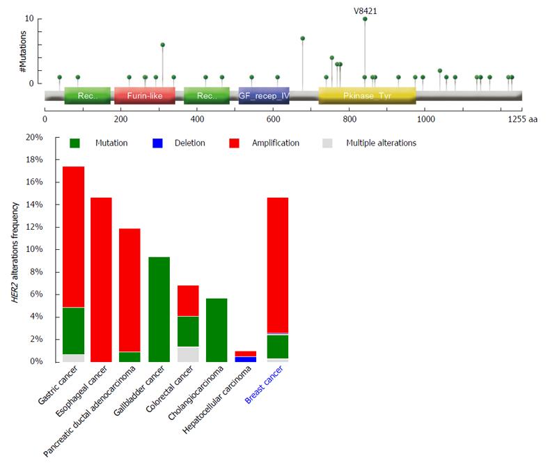
Figure 2 HER2 alterations frequencies from public datasets accessible from cBioPortal[16] in the most common tumors occurring in the digestive system compared to breast cancer.
Overall, HER2 amplifications occur more frequently in gastric, esophageal, and pancreatic cancers, whereas gallbladder cancer and cholangiocarcinoma characteristically show HER2 mutations (10% and 6% of cases, respectively). The domain structure of the erbB2 protein and gene alterations identified in primary carcinomas of the digestive system available from cBioPortal[16] (reported the top) show the presence of hotspot somatic mutations in the furin-like (binding site) and protein kinase domains. Mutation types are color-coded on the basis of the legend on the bottom right.
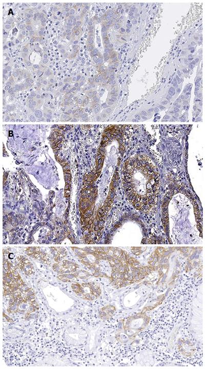
Figure 3 Representative micrographs of a gastric adenocarcinoma showing heterogeneous erbB2 immunohistochemical expression.
In this paradigmatic example of HER2 heterogeneity in gastric cancer, the tumor showed the coexistence of 2+ (A), 3+ (B, C), and negative areas (C), with the former immunohistochemical pattern involving the majority of the tumor cells. Original magnification × 200.
- Citation: Fusco N, Bosari S. HER2 aberrations and heterogeneity in cancers of the digestive system: Implications for pathologists and gastroenterologists. World J Gastroenterol 2016; 22(35): 7926-7937
- URL: https://www.wjgnet.com/1007-9327/full/v22/i35/7926.htm
- DOI: https://dx.doi.org/10.3748/wjg.v22.i35.7926


