Copyright
©The Author(s) 2016.
World J Gastroenterol. Sep 21, 2016; 22(35): 7926-7937
Published online Sep 21, 2016. doi: 10.3748/wjg.v22.i35.7926
Published online Sep 21, 2016. doi: 10.3748/wjg.v22.i35.7926
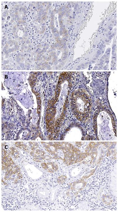
Figure 3 Representative micrographs of a gastric adenocarcinoma showing heterogeneous erbB2 immunohistochemical expression.
In this paradigmatic example of HER2 heterogeneity in gastric cancer, the tumor showed the coexistence of 2+ (A), 3+ (B, C), and negative areas (C), with the former immunohistochemical pattern involving the majority of the tumor cells. Original magnification × 200.
- Citation: Fusco N, Bosari S. HER2 aberrations and heterogeneity in cancers of the digestive system: Implications for pathologists and gastroenterologists. World J Gastroenterol 2016; 22(35): 7926-7937
- URL: https://www.wjgnet.com/1007-9327/full/v22/i35/7926.htm
- DOI: https://dx.doi.org/10.3748/wjg.v22.i35.7926


