Copyright
©The Author(s) 2016.
World J Gastroenterol. Sep 7, 2016; 22(33): 7536-7558
Published online Sep 7, 2016. doi: 10.3748/wjg.v22.i33.7536
Published online Sep 7, 2016. doi: 10.3748/wjg.v22.i33.7536
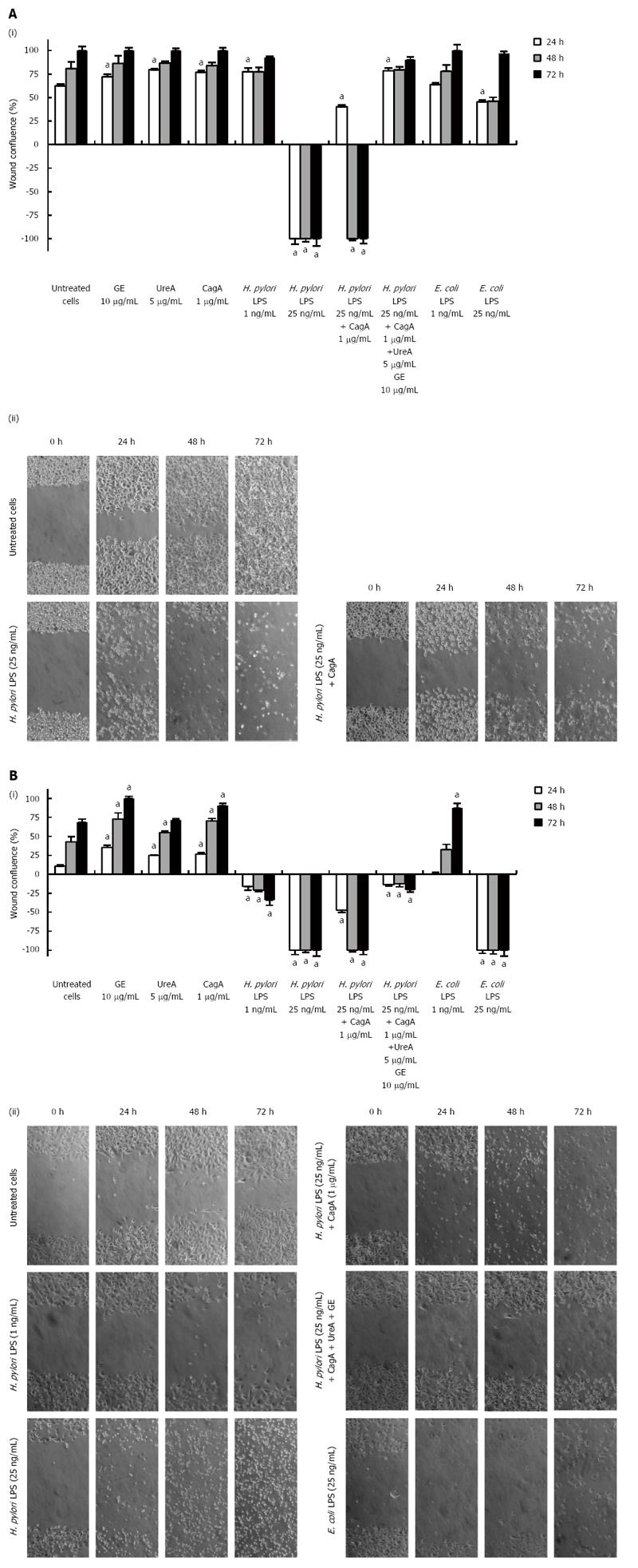
Figure 1 Migration effectiveness of human epithelial AGS cells and guinea pig fibroblasts assessed in a scratch assay.
(i) AGS cells (A) and fibroblasts (B) were grown to confluence and incubated overnight in RPMI-1640 medium/2% FBS/1% standard antibiotics. A wound was then made in a cell monolayer and culture medium alone or solutions of bacterial antigens were added. Wound areas were measured at 0, 24, 48 and 72 h after the challenge. Graphs of the average wound size against time, in which the results are shown for cells incubated alone (culture medium) or treated with GE (10 μg/mL), UreA (5 μg/mL), CagA (1 μg/mL) and Helicobacter pylori (H. pylori) LPS as well as Escherichia coli (E. coli) LPS (1 ng/mL or 25 ng/mL) or with a combination of H. pylori compounds: H. pylori LPS (25 ng/mL) and CagA (1 μg/mL) or H. pylori LPS (25 ng/mL), CagA (1 μg/mL), UreA (5 μg/mL) and GE (10 μg/mL). P = 0.03 vs untreated cells; (ii) Phase-contrast microscopy images were taken at the indicated time points and the extent of wound closure for each treatment variant was calculated as a percentage of migrating cells. Representative photos of each time point are shown (magnification × 200). aP = 0.03 vs untreated cells (according to the time of stimulation).
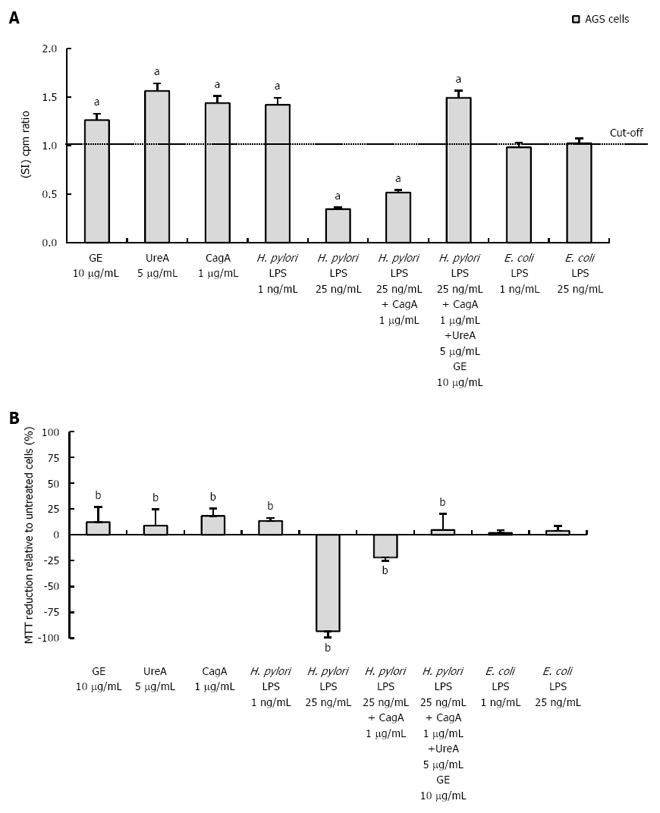
Figure 2 Influence of bacterial antigens on AGS cell proliferation and ability to reduce MTT.
A: The proliferative activity of AGS cells was estimated in cell cultures non-stimulated or stimulated for 24 h with bacterial antigens. After incubation, [3H]-thymidine incorporation into cellular DNA was analyzed. The graph shows the stimulation index (SI), which was calculated by dividing the radioactivity counts (cpm/min) for the cell cultures in the presence of a stimulus by the counts for control cell cultures in RPMI-1640 medium alone. The results are shown as SI ± SD of six independent experiments, performed in triplicates. aP = 0.03 vs untreated cells; B: AGS were treated for 24 h with bacterial antigens. After incubation, the ability of cells to reduce MTT was estimated. The graph shows the percentage of MTT reduction ± SD relative to untreated cells. The data represent the average values of four independent experiments performed in triplicates. The values have been normalized to those of the untreated cells. bP = 0.02 vs untreated cells.
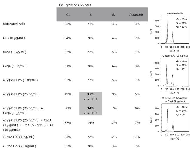
Figure 3 Effect of Helicobacter pylori antigens on the epithelial cell cycle profile.
AGS cells were incubated for 24 h in RPMI-1640 medium alone or in the presence of bacterial antigens. The cell cycle profile was determined by propidium iodide (PI) staining and the analysis was performed by flow cytometry. The data represent the percentage of cells in each cycle phase of six experiments. Statistically significant differences are indicated as P < 0.05 vs untreated cells and included in DNA histograms.
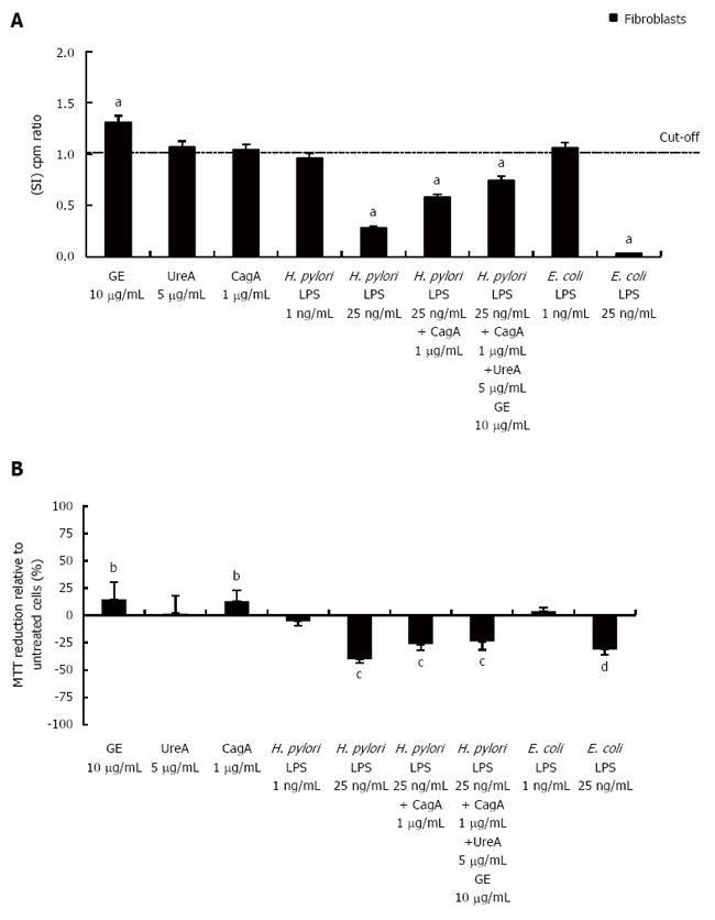
Figure 4 Influence of bacterial antigens on the ability of fibroblasts to proliferate and reduce MTT.
A: The proliferative activity of fibroblasts was estimated in cell cultures non-stimulated or stimulated for 24 h with bacterial antigens. After incubation, [3H]-thymidine incorporation into cellular DNA was analyzed. The graph shows the stimulation index (SI), which was calculated by dividing the radioactivity counts (cpm/min) for the cell cultures in the presence of a stimulus by the counts for control cell cultures in RPMI-1640 medium alone. The results are shown as SI ± SD of six independent experiments, performed in triplicates. aP = 0.02 vs untreated cells; B: Fibroblasts were treated for 24 h with bacterial antigens. After incubation, the ability of cells to reduce MTT was estimated. The graph shows the percentage of MTT reduction ± SD relative to untreated cells. The data represent the average values of four independent experiments performed in triplicates. The values have been normalized to those of the untreated cells. bP = 0.01; cP = 0.001; dP = 0.00002 vs untreated cells.
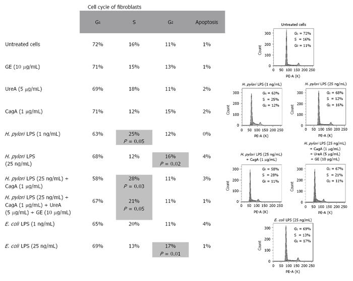
Figure 5 Effect of Helicobacter pylori antigens on the fibroblast cell cycle profile.
Fibroblasts were incubated for 24 h in RPMI-1640 medium alone or in the presence of bacterial antigens. The cell cycle profile was determined by propidium iodide (PI) staining and the analysis was performed by flow cytometry. The data represent the percentage of cells in each cycle phase of six experiments. Statistically significant differences are indicated as P < 0.05 vs untreated cells and included in DNA histograms.
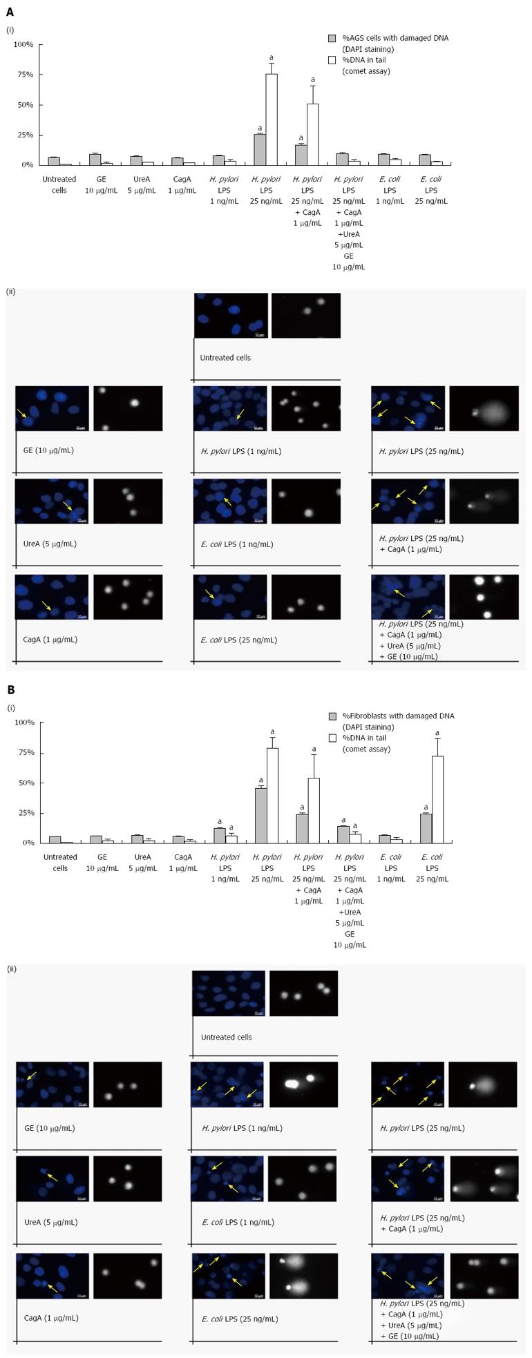
Figure 6 Genotoxic properties of Helicobacter pylori antigens assessed by 4’,6-diamidino-2-phenylindole staining and a comet assay.
The influence of Helicobacter pylori or E. coli antigens on DNA stability was estimated in AGS cells (A) and fibroblasts (B) 24 h after the challenge. A 4’,6-diamidino-2-phenylindole (DAPI) staining assay was used to visualize DNA changes in cell nuclei and a comet assay was applied to confirm DNA damage by the measurement of the percentage of DNA in the comet tail. Mean values were replicated of 50 comets each. The values are the means ± SD. aP < 0.05 vs untreated cells. (i) the graphs indicate the percentage of cells with DAPI stained nuclei (blue bars) and the percentage of nuclei with DNA in the comet tail (grey bars); and (ii) visualization of morphological changes in the cell nuclei after the treatment with bacterial antigens followed by DAPI staining and a comet assay. Imaging was performed using a fluorescent microscope (Axio Scope A1, Zeiss, Germany). The arrows indicate the damaged cell nuclei (magnification, × 1000). DNA tails were measured using the CASP software (latest beta version 1.2.3.beta2). Representative results of the comet assay were selected (magnification, × 400).
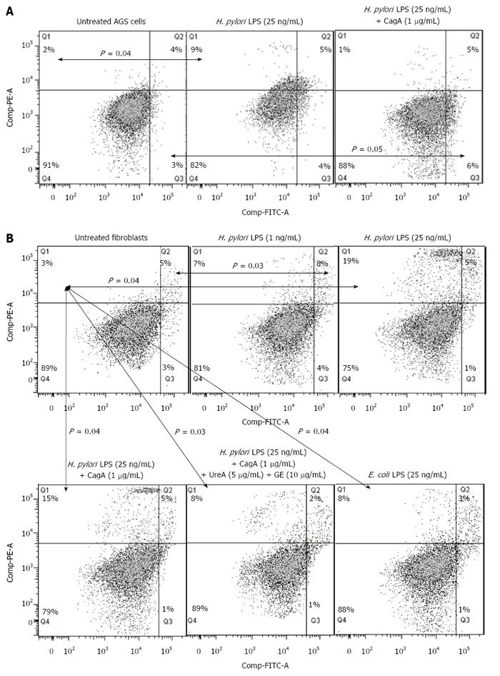
Figure 7 Type of cell death in response to Helicobacter pylori antigens.
Effects of bacterial antigens on AGS cells (A) and fibroblasts (B) concerning cell death were measured 24 h after the challenge by double staining of the cells with isothiocyanate fluorescein (FITC)-conjugated annexin V and propidium iodide (PI) using flow cytometry. Quadrants were designed as follows, Q4: Ann-V-/PI- - viable cells; Q3: Ann-V+/PI- - cells with the signs of early apoptosis; Q2: Ann-V+/PI+ - cells with the signs of late apoptosis; Q1: Ann-V-/PI+ - necrotic cells. All dot plots are a representation of equal cell populations (the fluorescence of 10000 cells was gated and counted using the FlowJo software). The data represent the average values of six independent experiments. Statistically significant differences are indicated as P < 0.05 vs untreated cells.
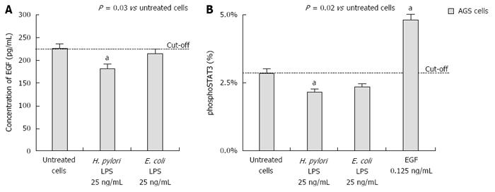
Figure 8 Helicobacter pylori lipopolysaccharide-driven inhibition of signal transducer and activator of transcription 3 phosphorylation and epidermal growth factor production.
A: The impact of Helicobacter pylori or E. coli lipopolysaccharide (LPS) on the secretion of epidermal growth factor (EGF) by AGS cells; B: the percentage of phospho-signal transducer and activator of transcription 3 (phosphoSTAT3) in AGS cells measured by cell-based ELISA. Cells treated with EGF (0.125 ng/mL) were used as a positive control. The cut-off value was related to the response of untreated cells. The data represent the average values of four independent experiments. Statistically significant differences are indicated as aP < 0.05 vs untreated cells.
- Citation: Mnich E, Kowalewicz-Kulbat M, Sicińska P, Hinc K, Obuchowski M, Gajewski A, Moran AP, Chmiela M. Impact of Helicobacter pylori on the healing process of the gastric barrier. World J Gastroenterol 2016; 22(33): 7536-7558
- URL: https://www.wjgnet.com/1007-9327/full/v22/i33/7536.htm
- DOI: https://dx.doi.org/10.3748/wjg.v22.i33.7536


