Copyright
©The Author(s) 2016.
World J Gastroenterol. Sep 7, 2016; 22(33): 7536-7558
Published online Sep 7, 2016. doi: 10.3748/wjg.v22.i33.7536
Published online Sep 7, 2016. doi: 10.3748/wjg.v22.i33.7536
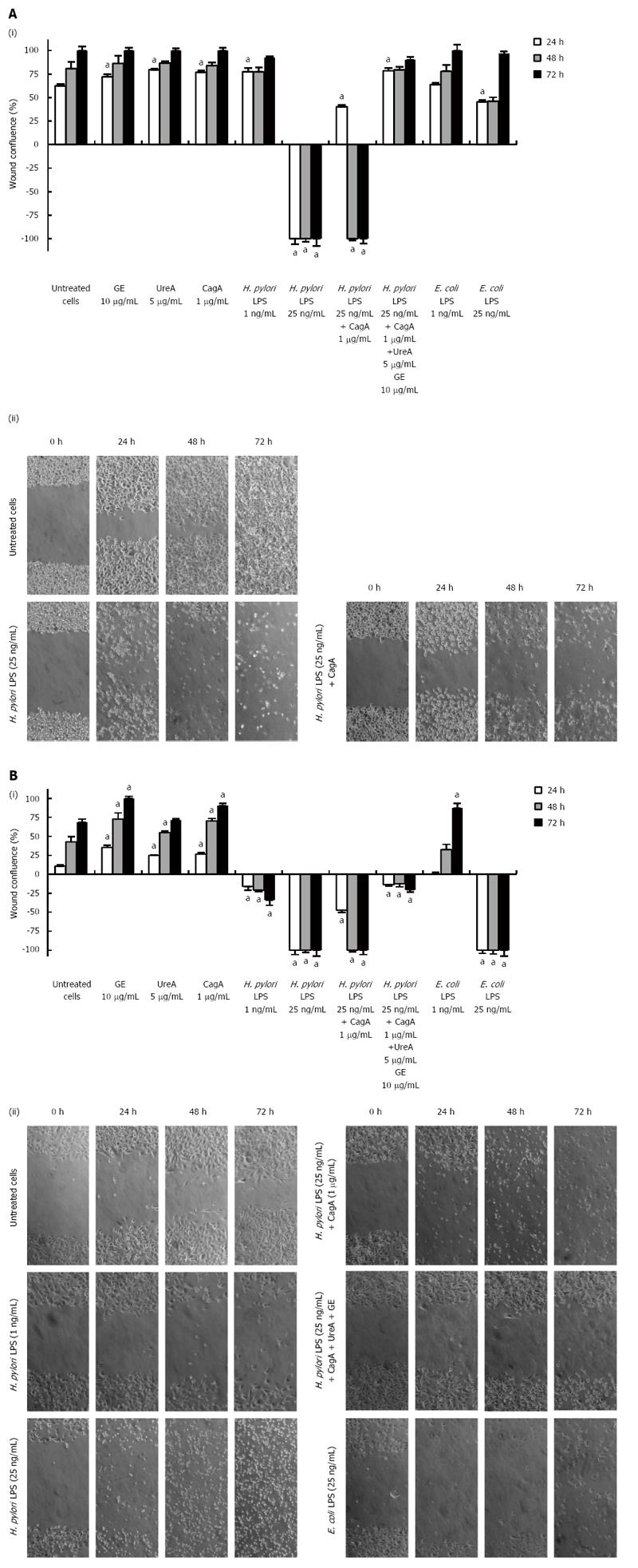
Figure 1 Migration effectiveness of human epithelial AGS cells and guinea pig fibroblasts assessed in a scratch assay.
(i) AGS cells (A) and fibroblasts (B) were grown to confluence and incubated overnight in RPMI-1640 medium/2% FBS/1% standard antibiotics. A wound was then made in a cell monolayer and culture medium alone or solutions of bacterial antigens were added. Wound areas were measured at 0, 24, 48 and 72 h after the challenge. Graphs of the average wound size against time, in which the results are shown for cells incubated alone (culture medium) or treated with GE (10 μg/mL), UreA (5 μg/mL), CagA (1 μg/mL) and Helicobacter pylori (H. pylori) LPS as well as Escherichia coli (E. coli) LPS (1 ng/mL or 25 ng/mL) or with a combination of H. pylori compounds: H. pylori LPS (25 ng/mL) and CagA (1 μg/mL) or H. pylori LPS (25 ng/mL), CagA (1 μg/mL), UreA (5 μg/mL) and GE (10 μg/mL). P = 0.03 vs untreated cells; (ii) Phase-contrast microscopy images were taken at the indicated time points and the extent of wound closure for each treatment variant was calculated as a percentage of migrating cells. Representative photos of each time point are shown (magnification × 200). aP = 0.03 vs untreated cells (according to the time of stimulation).
- Citation: Mnich E, Kowalewicz-Kulbat M, Sicińska P, Hinc K, Obuchowski M, Gajewski A, Moran AP, Chmiela M. Impact of Helicobacter pylori on the healing process of the gastric barrier. World J Gastroenterol 2016; 22(33): 7536-7558
- URL: https://www.wjgnet.com/1007-9327/full/v22/i33/7536.htm
- DOI: https://dx.doi.org/10.3748/wjg.v22.i33.7536


