Copyright
©The Author(s) 2016.
World J Gastroenterol. Sep 7, 2016; 22(33): 7536-7558
Published online Sep 7, 2016. doi: 10.3748/wjg.v22.i33.7536
Published online Sep 7, 2016. doi: 10.3748/wjg.v22.i33.7536
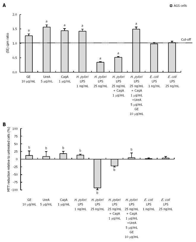
Figure 2 Influence of bacterial antigens on AGS cell proliferation and ability to reduce MTT.
A: The proliferative activity of AGS cells was estimated in cell cultures non-stimulated or stimulated for 24 h with bacterial antigens. After incubation, [3H]-thymidine incorporation into cellular DNA was analyzed. The graph shows the stimulation index (SI), which was calculated by dividing the radioactivity counts (cpm/min) for the cell cultures in the presence of a stimulus by the counts for control cell cultures in RPMI-1640 medium alone. The results are shown as SI ± SD of six independent experiments, performed in triplicates. aP = 0.03 vs untreated cells; B: AGS were treated for 24 h with bacterial antigens. After incubation, the ability of cells to reduce MTT was estimated. The graph shows the percentage of MTT reduction ± SD relative to untreated cells. The data represent the average values of four independent experiments performed in triplicates. The values have been normalized to those of the untreated cells. bP = 0.02 vs untreated cells.
- Citation: Mnich E, Kowalewicz-Kulbat M, Sicińska P, Hinc K, Obuchowski M, Gajewski A, Moran AP, Chmiela M. Impact of Helicobacter pylori on the healing process of the gastric barrier. World J Gastroenterol 2016; 22(33): 7536-7558
- URL: https://www.wjgnet.com/1007-9327/full/v22/i33/7536.htm
- DOI: https://dx.doi.org/10.3748/wjg.v22.i33.7536


