Copyright
©The Author(s) 2016.
World J Gastroenterol. Aug 14, 2016; 22(30): 6960-6964
Published online Aug 14, 2016. doi: 10.3748/wjg.v22.i30.6960
Published online Aug 14, 2016. doi: 10.3748/wjg.v22.i30.6960
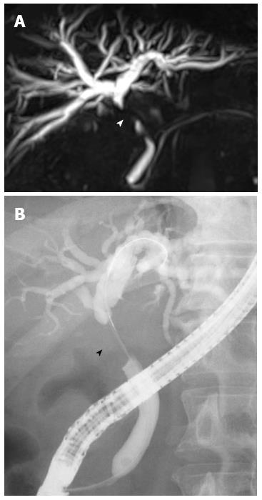
Figure 1 Magnetic resonance cholangiopancreatography (A) and endoscopic retrograde cholangiopancreatography (B) of the bile duct.
Severe stenosis of the middle common bile duct was observed (arrowhead).
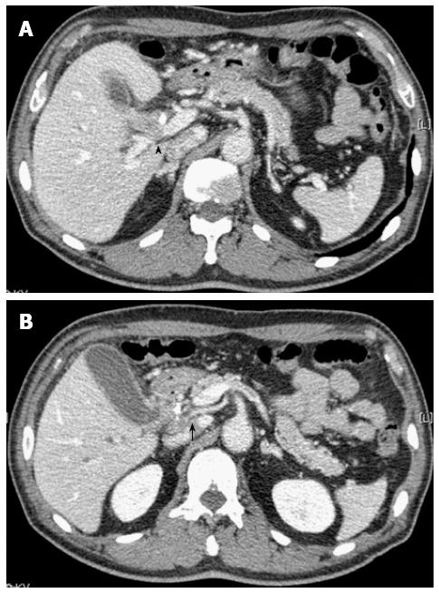
Figure 2 Abdominal computed tomography images at two different levels are shown.
A: Computed tomography (CT) showed a low-density mass measuring 18 mm in diameter located in the middle bile duct adjacent to the portal vein (arrowhead). An endoscopic nasobiliary drainage tube was observed in the bile duct; B: CT showed a low-density mass involving the right hepatic artery from the superior mesenteric artery in the middle bile duct (arrow).
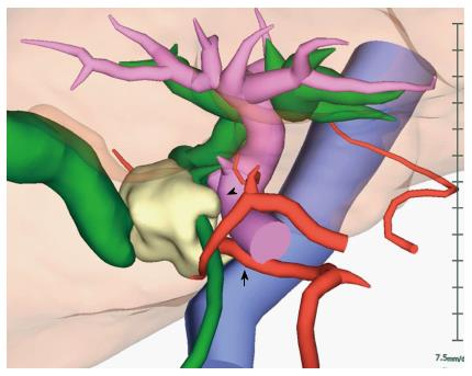
Figure 3 3D reconstructed computed tomography fused with an magnetic resonance cholangiopancreatography image.
The tumor was located in the middle bile duct. The tumor was suspected to have invaded the portal vein (arrowhead) and the right hepatic artery from the superior mesenteric artery (arrow).
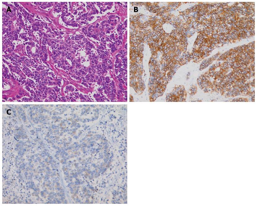
Figure 4 Histopathologic appearance of the neuroendocrine carcinoma.
A: The cells were round or oval, hyperchromatic, and had an increased nucleus-to-cytoplasm ratio (hematoxylin-eosin staining, magnification × 200); B: Immunohistochemically, tumor cells were diffusely positive for CD56, a membrane protein usually present in neuroendocrine cells; C: Immunohistochemically, tumor cells were positive for synaptophysin, which is typically expressed on the surface of neurons or endothelial cells.
- Citation: Oshiro Y, Gen R, Hashimoto S, Oda T, Sato T, Ohkohchi N. Neuroendocrine carcinoma of the extrahepatic bile duct: A case report. World J Gastroenterol 2016; 22(30): 6960-6964
- URL: https://www.wjgnet.com/1007-9327/full/v22/i30/6960.htm
- DOI: https://dx.doi.org/10.3748/wjg.v22.i30.6960


