Copyright
©The Author(s) 2016.
World J Gastroenterol. Aug 14, 2016; 22(30): 6955-6959
Published online Aug 14, 2016. doi: 10.3748/wjg.v22.i30.6955
Published online Aug 14, 2016. doi: 10.3748/wjg.v22.i30.6955
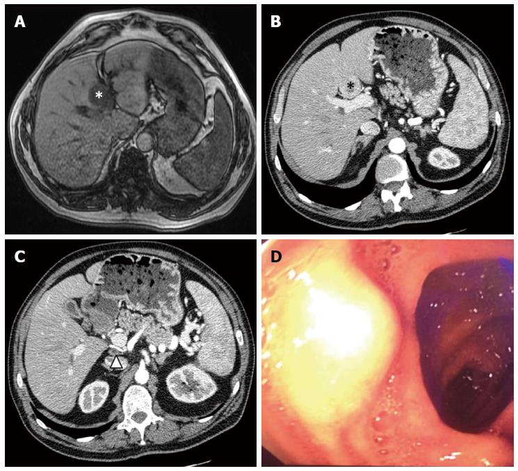
Figure 1 Pre-operative imaging.
A: Pre-operative MRI of the liver, showing liver metastases of gastrinoma (white star); B: Pre-operative CT scan of the abdomen, showing liver metastases of gastrinoma (black star); C: Pre-operative CT scan of the abdomen, showing the retroportal lymph node (black triangle); D: Upper endoscopy, showing the tumor on the superior duodenal flexure. MRI: Magnetic resonance imaging; CT: Computed tomography.
Figure 2 Intraoperative findings.
A: Duodenotomy, showing the gastrinoma on the superior duodenal flexure (white triangle), next to the pancreatic head (black star); B: The retroportal lymph node; C: Hepatotomy during left hepatectomy (1: Common bile duct; 2: Left hepatic duct, 3: Right hepatic duct, 4: Right posterior segmental duct, 5: Cystic duct).
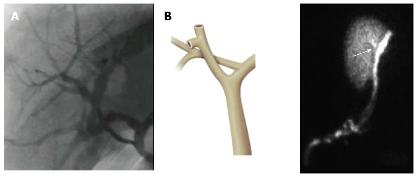
Figure 3 Intraoperative cholangiography findings.
A: Intraoperative cholangiography, showing dilatation of the right posterior segmental duct and its confluence with the left hepatic duct; B: A frontal view of the anatomic variation of the right posterior segmental duct and its junction with the left hepatic duct.
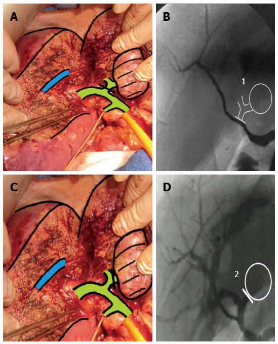
Figure 4 Correspondence between anatomic and radiologic features during left hepatectomy.
A: Clamping downstream of the confluence of right posterior segmental duct with the left hepatic duct; B: Intraoperative cholangiography, showing the absence of opacification of the right posterior segmental duct; C: Clamping upstream of the confluence of the right posterior segmental duct with the left hepatic duct; D: Intraoperative cholangiography, showing opacification of the right posterior segmental duct.
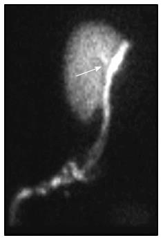
Figure 5 Liver scintigraphy, showing the biliary fistula (white arrow).
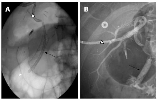
Figure 6 Rendezvous procedure.
A: X-ray of the abdomen, showing catheterization of right posterior segmental duct and the common bile duct with external drainage (white triangle), the position of the covered biliary stent (black arrow) and the position of the covered duodenal stent (white arrow) next to the distal end of the biliary stent; B: Opacification of external drainage (white triangle), showing the continuity between the right posterior segmental duct and the common bile duct provided by the biliary stent (black arrow).
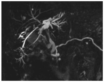
Figure 7 Magnetic resonance image of the liver 1 year after removal of the stents, showing moderate dilatation of right posterior segmental duct and continuity of the hepatic ducts.
- Citation: Gracient A, Rebibo L, Delcenserie R, Yzet T, Regimbeau JM. Combined radiologic and endoscopic treatment (using the “rendezvous technique”) of a biliary fistula following left hepatectomy. World J Gastroenterol 2016; 22(30): 6955-6959
- URL: https://www.wjgnet.com/1007-9327/full/v22/i30/6955.htm
- DOI: https://dx.doi.org/10.3748/wjg.v22.i30.6955


