Copyright
©The Author(s) 2016.
World J Gastroenterol. Aug 14, 2016; 22(30): 6955-6959
Published online Aug 14, 2016. doi: 10.3748/wjg.v22.i30.6955
Published online Aug 14, 2016. doi: 10.3748/wjg.v22.i30.6955
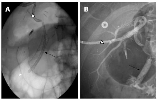
Figure 6 Rendezvous procedure.
A: X-ray of the abdomen, showing catheterization of right posterior segmental duct and the common bile duct with external drainage (white triangle), the position of the covered biliary stent (black arrow) and the position of the covered duodenal stent (white arrow) next to the distal end of the biliary stent; B: Opacification of external drainage (white triangle), showing the continuity between the right posterior segmental duct and the common bile duct provided by the biliary stent (black arrow).
- Citation: Gracient A, Rebibo L, Delcenserie R, Yzet T, Regimbeau JM. Combined radiologic and endoscopic treatment (using the “rendezvous technique”) of a biliary fistula following left hepatectomy. World J Gastroenterol 2016; 22(30): 6955-6959
- URL: https://www.wjgnet.com/1007-9327/full/v22/i30/6955.htm
- DOI: https://dx.doi.org/10.3748/wjg.v22.i30.6955


