Copyright
©The Author(s) 2016.
World J Gastroenterol. Mar 21, 2016; 22(11): 3289-3295
Published online Mar 21, 2016. doi: 10.3748/wjg.v22.i11.3289
Published online Mar 21, 2016. doi: 10.3748/wjg.v22.i11.3289
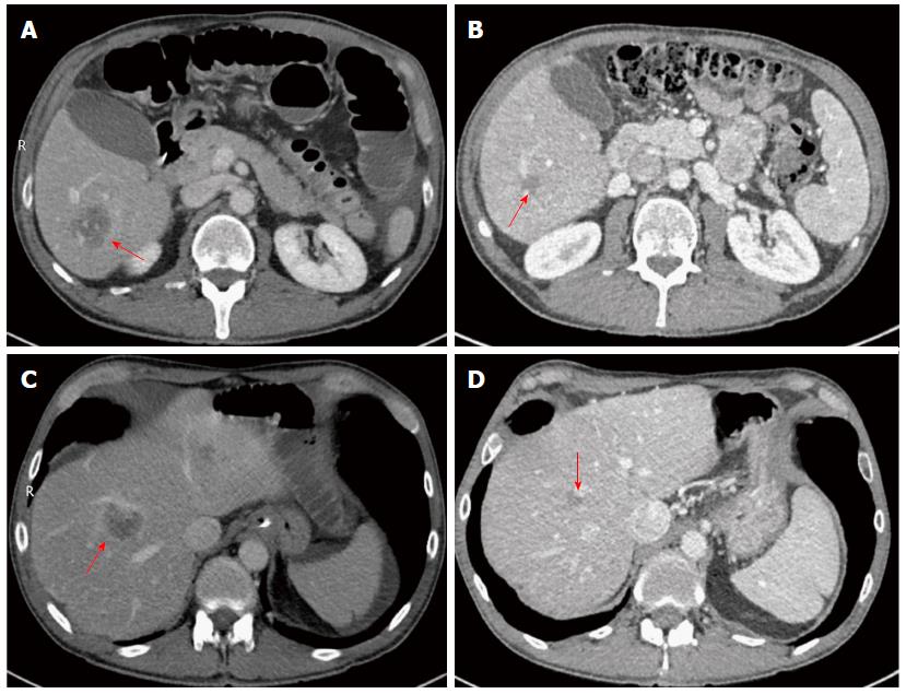
Figure 1 Computed tomography imaging of liver.
Evidence of metastatic lesions in the right hemi-liver segments VI (A) and VIII (C) pre-chemotherapy. Two residual lesions in the right hemi-liver segments VI (B) and VIII (D) post-chemotherapy. Red arrows indicate metastatic lesions.
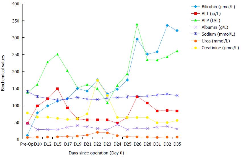
Figure 2 Trend in biochemical markers following hepatectomy.
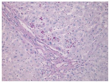
Figure 3 Histopathological findings.
Alpha-1-antitrypsin globules within peri-portal hepatocytes (diastase periodic acid schiff, magnification × 200).
- Citation: Norton B, Denson J, Briggs C, Bowles M, Stell D, Aroori S. Delayed diagnosis of alpha-1-antitrypsin deficiency following post-hepatectomy liver failure: A case report. World J Gastroenterol 2016; 22(11): 3289-3295
- URL: https://www.wjgnet.com/1007-9327/full/v22/i11/3289.htm
- DOI: https://dx.doi.org/10.3748/wjg.v22.i11.3289


