Copyright
©The Author(s) 2016.
World J Gastroenterol. Mar 21, 2016; 22(11): 3285-3288
Published online Mar 21, 2016. doi: 10.3748/wjg.v22.i11.3285
Published online Mar 21, 2016. doi: 10.3748/wjg.v22.i11.3285
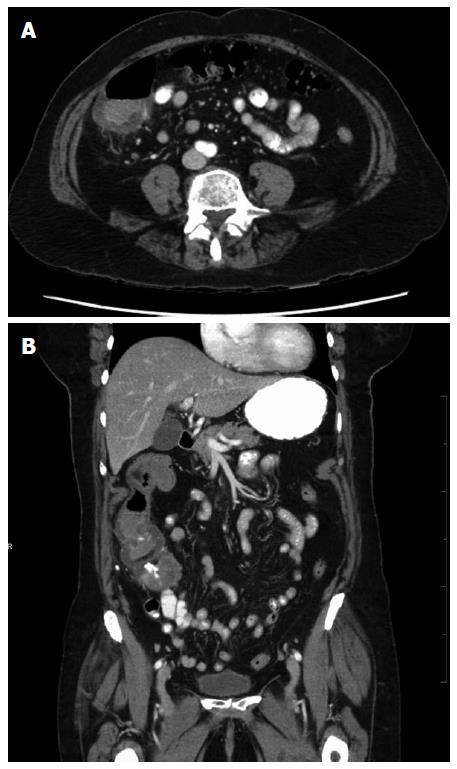
Figure 1 Computed tomographic scan images of contained colonic perforation suspected secondary to cecal retroflexion.
The cecum with an associated contained fluid collection is shown in axial (A) and coronal (B) views.
- Citation: Geng Z, Agrawal D, Singal AG, Kircher S, Gupta S. Contained colonic perforation due to cecal retroflexion. World J Gastroenterol 2016; 22(11): 3285-3288
- URL: https://www.wjgnet.com/1007-9327/full/v22/i11/3285.htm
- DOI: https://dx.doi.org/10.3748/wjg.v22.i11.3285


