Copyright
©The Author(s) 2015.
World J Gastroenterol. Mar 7, 2015; 21(9): 2651-2657
Published online Mar 7, 2015. doi: 10.3748/wjg.v21.i9.2651
Published online Mar 7, 2015. doi: 10.3748/wjg.v21.i9.2651
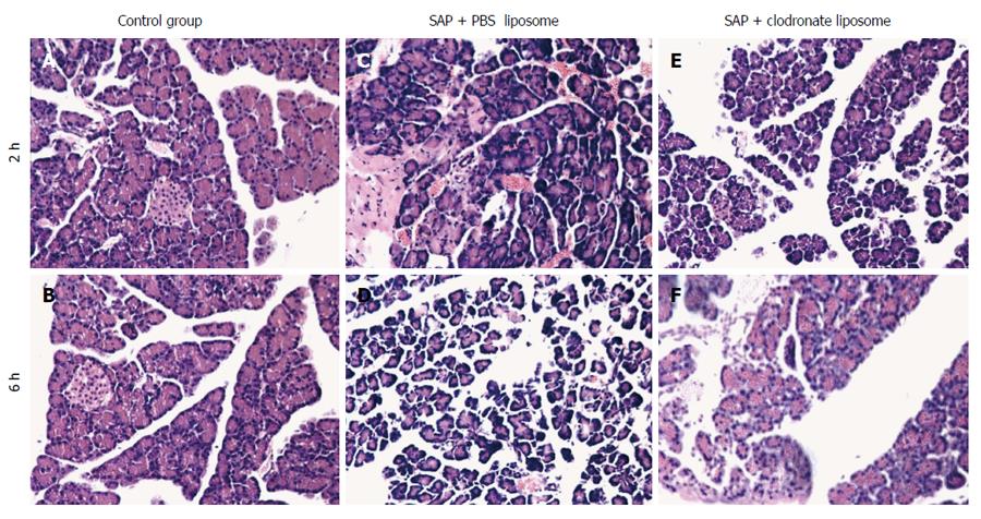
Figure 1 Pathological changes in the pancreas.
The pancreas of rats in the C group showed no morphological changes (A, B); Significant inflammatory cell infiltration was observed in the P group (C, D); Mild pancreatic edema, hemorrhage, and inflammatory cell infiltration were observed in the T group (E, F). Original magnification: × 200. SAP: Severe acute pancreatitis; PBS: Phosphate-buffered saline.
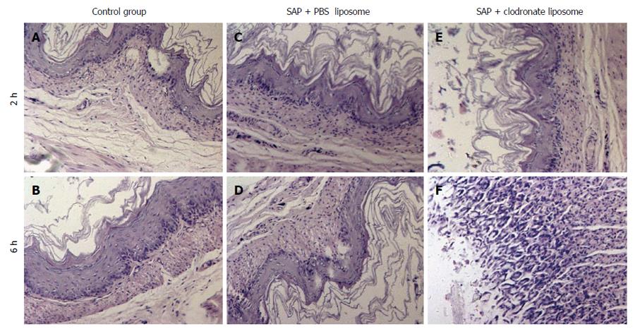
Figure 2 Morphological changes in the gastric mucosa.
Gastric sections with normal mucosa, histopathologically graded as 0-1 (A, B); Gastric sections with mucosa necrosis, histopathologically graded as 3-4 (C, D); Slightly damaged gastric mucosa was observed in the group treated with drug-containing liposomes (E, F). Original magnification: × 200. SAP: Severe acute pancreatitis; PBS: Phosphate-buffered saline.
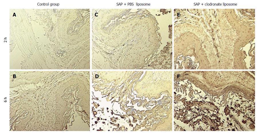
Figure 3 Terminal deoxynucleotidyl transferase dUTP nick end labeling analysis of apoptosis in each group.
Apoptosis was not observed in the gastric mucosa of the C group (A, B); The apoptotic cell indices of the mucosal cells were higher in the T group (C, D) than in the P group (E, F), 2 and 6 h after induction of severe acute pancreatitis. Original magnification: × 200. SAP: Severe acute pancreatitis; PBS: Phosphate-buffered saline.
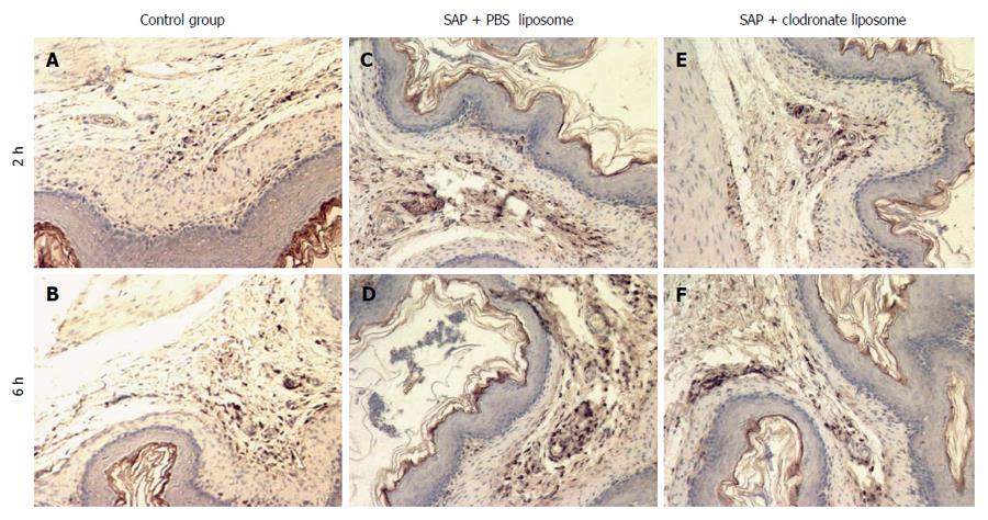
Figure 4 Number of macrophages observed in the gastric mucosa.
Macrophages were detected immunohistochemically using anti-CD68 antibodies. CD68-positive cells were rarely seen in the control rats (A, B); Numerous CD68-positive cell clusters were observed in the P group (C, D); Fewer CD68-positive cells were observed in the T group compared with the P group (E, F). Original magnification: × 200. SAP: Severe acute pancreatitis; PBS: Phosphate-buffered saline.
- Citation: Dang SC, Wang H, Zhang JX, Cui L, Jiang DL, Chen RF, Qu JG, Shen XQ, Chen M, Gu M. Are gastric mucosal macrophages responsible for gastric injury in acute pancreatitis? World J Gastroenterol 2015; 21(9): 2651-2657
- URL: https://www.wjgnet.com/1007-9327/full/v21/i9/2651.htm
- DOI: https://dx.doi.org/10.3748/wjg.v21.i9.2651


