Copyright
©The Author(s) 2015.
World J Gastroenterol. Feb 21, 2015; 21(7): 2260-2262
Published online Feb 21, 2015. doi: 10.3748/wjg.v21.i7.2260
Published online Feb 21, 2015. doi: 10.3748/wjg.v21.i7.2260
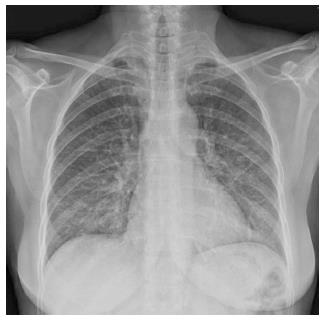
Figure 1 Chest X-ray at hospital admission in the Pneumology Department showed a left predominant interstitial pattern.
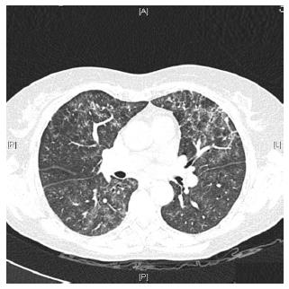
Figure 2 High-resolution computed tomography showed areas of diffuse ground glass opacities and cylindrical bronchiectasis in both lungs.
A: Anterior; R: Right; L: Left; P: Posterior.
- Citation: Casanova MJ, Chaparro M, Valenzuela C, Cisneros C, Gisbert JP. Adalimumab-induced interstitial pneumonia in a patient with Crohn’s disease. World J Gastroenterol 2015; 21(7): 2260-2262
- URL: https://www.wjgnet.com/1007-9327/full/v21/i7/2260.htm
- DOI: https://dx.doi.org/10.3748/wjg.v21.i7.2260


