Copyright
©The Author(s) 2015.
World J Gastroenterol. Feb 7, 2015; 21(5): 1663-1665
Published online Feb 7, 2015. doi: 10.3748/wjg.v21.i5.1663
Published online Feb 7, 2015. doi: 10.3748/wjg.v21.i5.1663
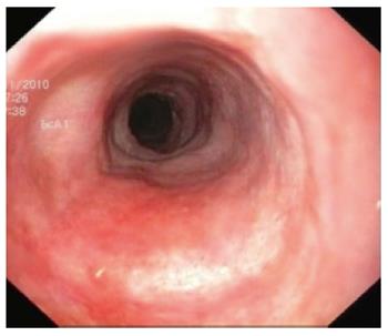
Figure 1 Endoscopic view of the esophagus showing granular changes of the inner surface as well as whitish discoloration.
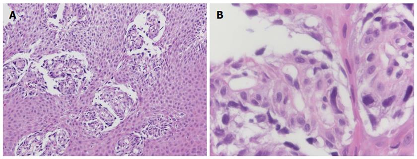
Figure 2 Initial forceps biopsy results.
A: Tangential section through the esophageal epithelium. Numerous atypical cells adjacent to the subepithelial papillae, overlying squamous epithelium without atypia. Hematoxylin and eosin stain; original magnification × 10; B: High-magnification of tumor cells with highly atypical nuclei, loss of cell cohesion, and either polygonal or spindle-cell shapes.
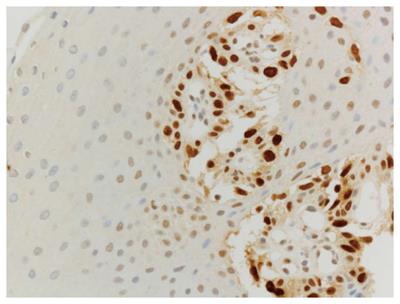
Figure 3 Esophageal squamous epithelium showing atypical cells with accumulation of the p53-protein, restricted to the basal portion of the epithelium.
- Citation: Sarbia M, Wolfer S, Karimi D, Eimiller A. High-grade dysplasia, restricted to the basal cell layer involving the entire esophagus. World J Gastroenterol 2015; 21(5): 1663-1665
- URL: https://www.wjgnet.com/1007-9327/full/v21/i5/1663.htm
- DOI: https://dx.doi.org/10.3748/wjg.v21.i5.1663


