Copyright
©The Author(s) 2015.
World J Gastroenterol. Nov 28, 2015; 21(44): 12519-12543
Published online Nov 28, 2015. doi: 10.3748/wjg.v21.i44.12519
Published online Nov 28, 2015. doi: 10.3748/wjg.v21.i44.12519
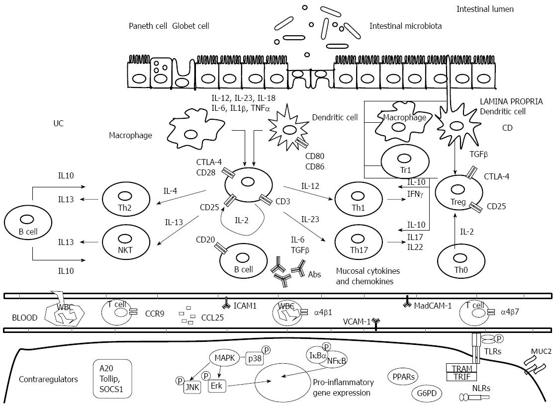
Figure 1 Inflammatory and regulatory pathways involved in inflammatory bowel disease pathogenesis.
Crohn’s disease (CD) is characterized by the generation of Th1- and Th17 T cell responses driven by the production of interleukin (IL)-12, IL-18, IL-23, IL-6 and tumor necrosis factor (TNF)-α by dendritic cells and macrophages. Th1-cells secrete IL-2, IL-17, interferon (IFN)-γ, and TNF-α. Ulcerative colitis (UC) is characterized by a Th2- T cell, and NKT response mediated by IL-5 and IL-13. T cell responses initiate an inflammatory cascade that involves endothelial activation, chemokine production, and white blood cell recruitment. Inappropriate triggering and maintenance of these pathogenic responses has been associated with innate immunity defects, e.g., lack of efficient control by anti-inflammatory cytokines such as IL-10 and transforming growth factor (TGF)-β. In the bottom of the figure, intracellular markers of activation are represented. Ab: antibody; CTLA-4: Cytotoxic T lymphocyte antigen-4; ICAM-1: Intercellular adhesion molecule-1; MAdCAM-1: Mucosal addressin cell adhesion molecule-1; VCAM-1: Vascular cell adhesion molecule-1; TLRs: Toll-like receptors; NLR: NOD-like receptor; NKT: Natural killer T.
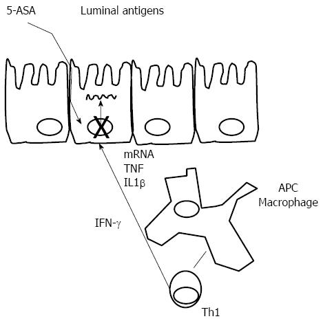
Figure 2 Mesalazine mode of action.
Antigen presenting cells (APCs) recognize luminal antigens penetrating the colonic wall and through interaction with Th1 cells induce the production of interferon (IFN)-γ. IFN-γ activates epithelial cells. 5-aminosalicylic acid (5-ASA) is able to block transcription of inflammatory cytokines in colonic epithelial cells. IL: Interleukin; TNF: Tumor necrosis factor.
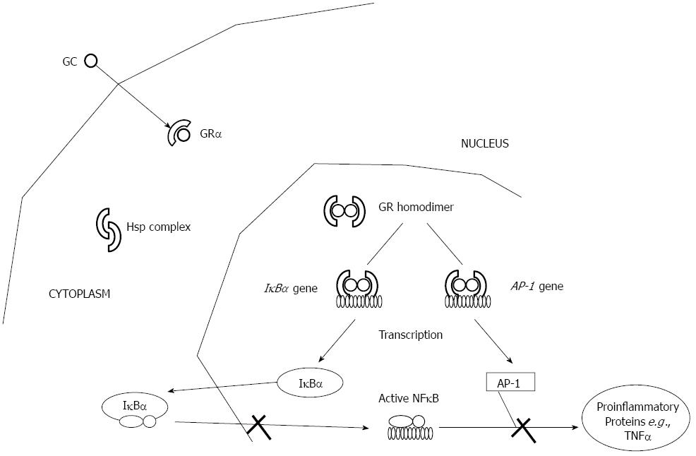
Figure 3 Glucocorticoids mode of action.
AP: Activator protein; GC: Glucocorticoids; GR: Glucocorticoid receptor; TNF: Tumor necrosis factor; NF: Nuclear factor.
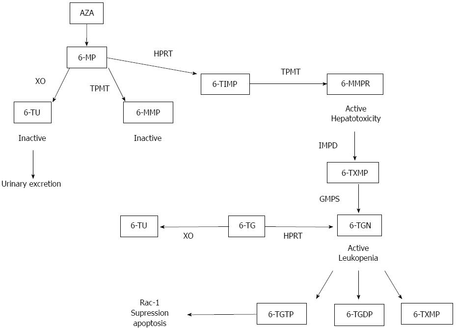
Figure 4 Thiopurines metabolic pathway.
XO: Xanthin oxidase; 6-TU: 6-thiouracil; TPMT: Thiopurine methyltransferase; HPRT: Hypoxanthine phosporybosil transferase; 6-MMP: 6-methyl mercaptopurine; 6-TIMP: 6-thiosine 5’ monophosphate; 6-MMPR: 6-methyl mercaptopurine ribonucleotide; IMPD: Inosine monophosphate dehydrogenase; 6-TXMP: 6-Thioxanthosine monophosphate; GMPS: Guanosine monophosphate synthetase; 6-TGN: 6-thioguanine nucleotide; 6-TG: 6-thioguanine; 6-TGDP: 6-thioguanine diphosphate; 6-TGTP: 6-thioguanine triphosphate.
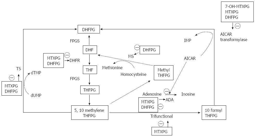
Figure 5 Methotrexate mode of action.
DHF: Dihydrofolate; FPGS: Folil polyglutamate synthetase; DHFPG: Dihydrofolate polyglutamates; DHFR: Dihydrofolate reductase; THF: Tetrahydrofolate; THFPG: Tetrahydrofolate polyglutamates; TS: Thymidylate synthetase; MS: Methionine synthetase; ADA: Adenosine deaminase; IMP: Inosine monophosphate; AICAR: 5-aminoimidazole-4-carbox-amide ribonucleotide.
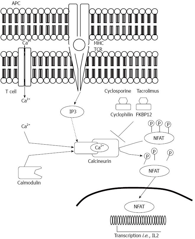
Figure 6 Mode of action of calcineurin inhibitors.
APC: Antigen presenting cell; MHC: Major histocompatibility complex; NFAT: Nuclear factor of activated T cells; TCR: T cell receptor; IP3: Inositol triphosphate.
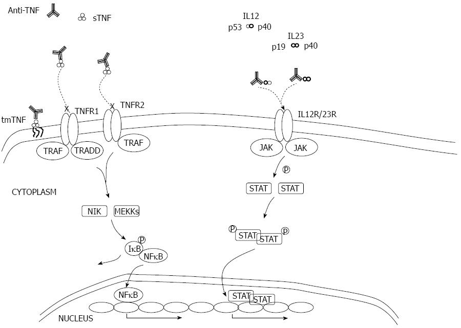
Figure 7 Mode of action of anti- tumor necrosis factor α and anti-IL-12/23.
TNF: Tumor necrosis factor; tmTNF: Transmembrane TNF; sTNF: Soluble TNF; TRAF/TRADD: TLRs adaptors; JAK: Janus kinase; STAT: Signal transducer and activator of transcription.
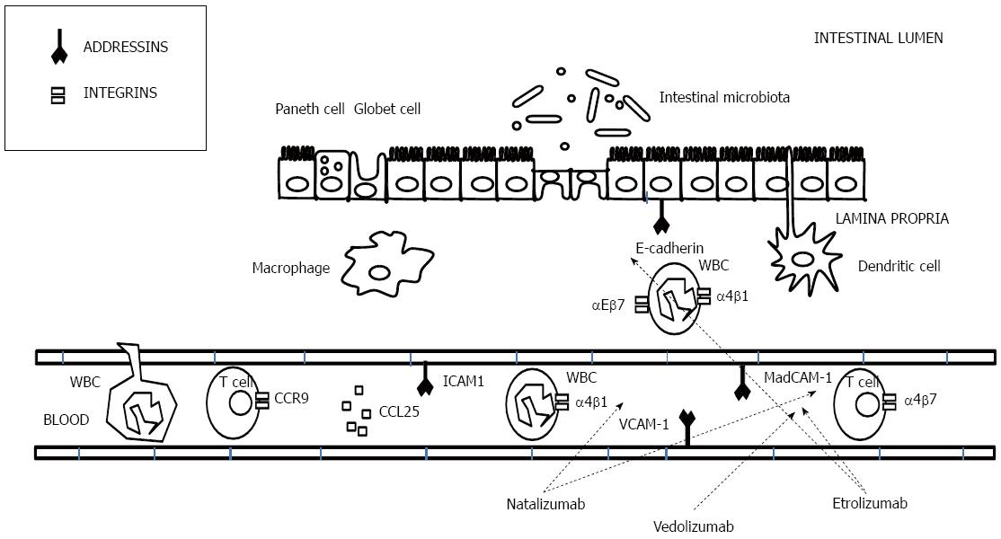
Figure 8 Mechanism of action of anti-adhesion drugs.
- Citation: Quetglas EG, Mujagic Z, Wigge S, Keszthelyi D, Wachten S, Masclee A, Reinisch W. Update on pathogenesis and predictors of response of therapeutic strategies used in inflammatory bowel disease. World J Gastroenterol 2015; 21(44): 12519-12543
- URL: https://www.wjgnet.com/1007-9327/full/v21/i44/12519.htm
- DOI: https://dx.doi.org/10.3748/wjg.v21.i44.12519


