Copyright
©The Author(s) 2015.
World J Gastroenterol. Nov 21, 2015; 21(43): 12392-12402
Published online Nov 21, 2015. doi: 10.3748/wjg.v21.i43.12392
Published online Nov 21, 2015. doi: 10.3748/wjg.v21.i43.12392
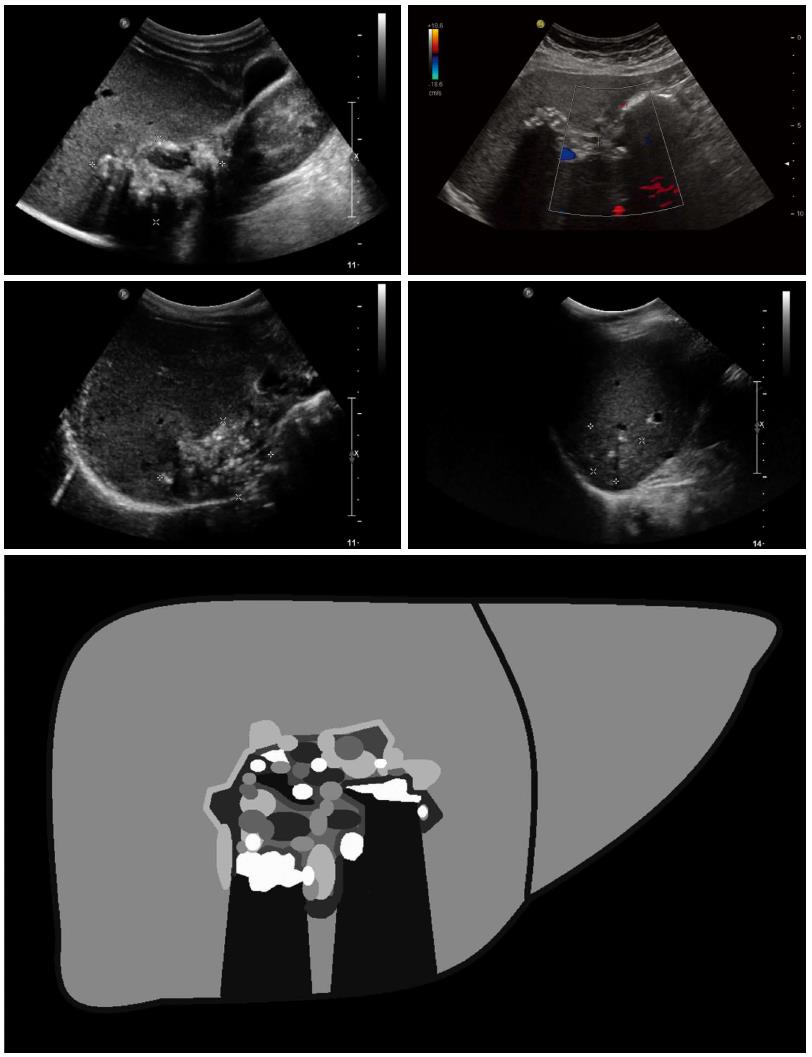
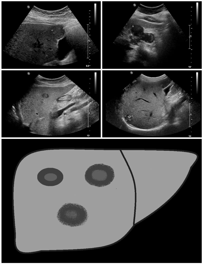
Figure 1 Hailstorm: The typical hailstorm appearance is characterized by indistinct, irregular boundaries, non-homogeneous pattern and hyperechoic formations, with or without dorsal acoustic shadow.
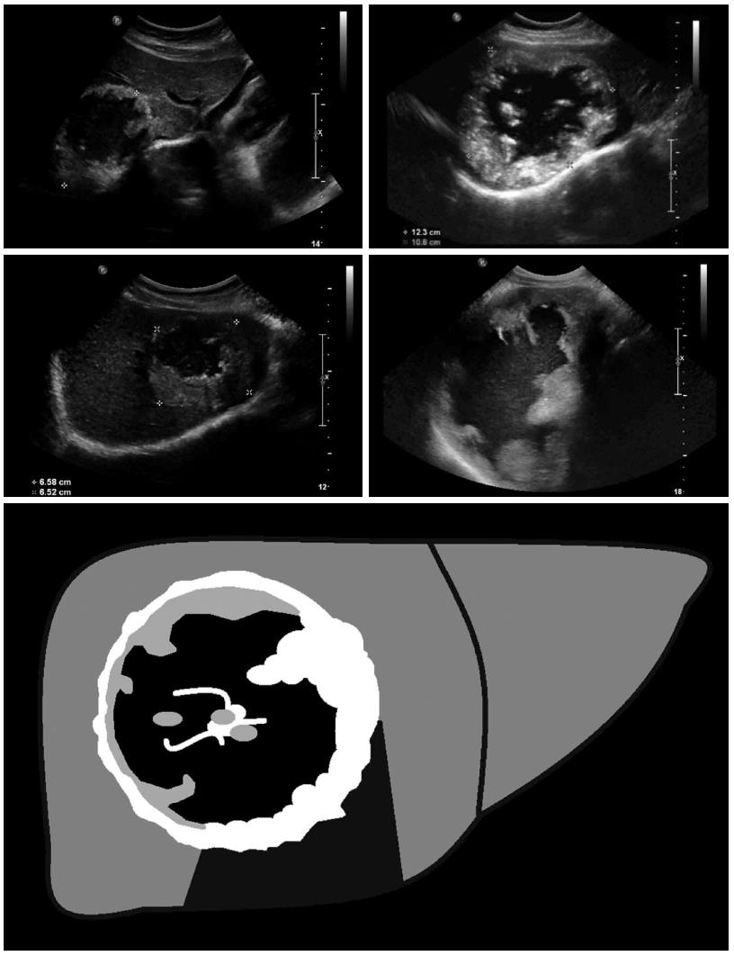
Figure 2 Pseudocystic: Pseudocystic alveolar echinococcosis lesions are primarily characterized by an hyperechoic, irregular and non-homogeneous rim that is non-vascularized at power Doppler and color-coded duplex ultrasonography.
It may appear to be > 10 mm in thickness. There is a hypo- or anechoic, often non-homogeneous central zone that may contain hyperechoic material. Pseudocystic lesions may be already present at first diagnosis and involve an entire hepatic lobe, or may develop from primary hailstorm lesions following therapy with benzimidazoles.
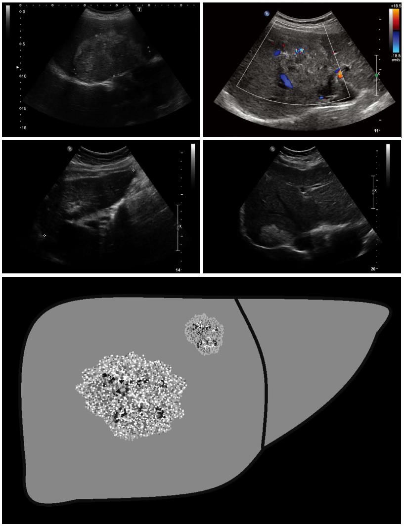
Figure 3 Hemangioma-like: These lesions are difficult to distinguish from atypical (e.
g., partially thrombosed) hemangiomas, and often represent a significant diagnostic challenge. Sonomorphologically, the lesions present as a relatively clearly demarcated non-homogeneous tumor that appears hyperechoic in comparison with the surrounding hepatic parenchyma. Echogenicity ranges from slightly and non-homogeneously hyperechoic to strongly and homogeneous hyperechoic.
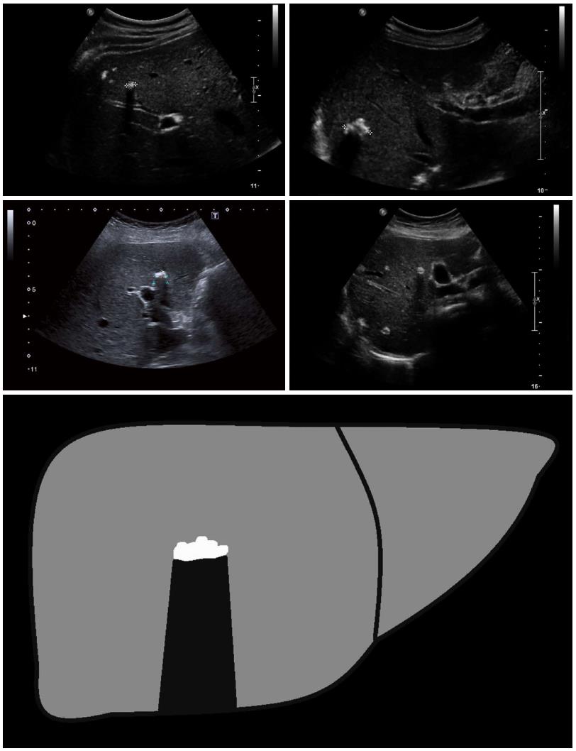
Figure 4 Ossification: The ossification pattern presents with solitary or grouped, mostly sharply delineated lesions with dorsal acoustic shadow.
In terms of their differential diagnosis, these alveolar echinococcosis (AE) lesions are often difficult to distinguish from inflammatory or hyperechoic metastases of various carcinomas. Very large ossification-type AE lesions represent a rarity. Both uni- and multifocal involvement is possible.
Figure 5 Metastasis-like: Beside the hemangioma-like lesions, the metastasis-like lesions of alveolar echinococcosis represent the greatest diagnostic challenge.
Mostly hypoechoic, these lesions exhibit as a typical characteristic-compared to typical hepatic metastases (e.g., of colorectal cancer)-the absence of the halo phenomenon. Instead, there is a central, hyperechoic, non-homogeneous scar.
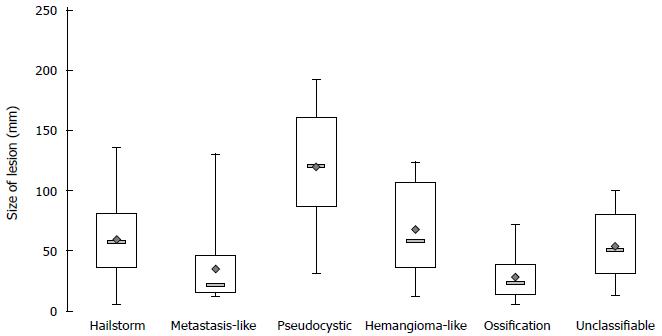
Figure 6 Lesion size depending on the sonomorphological pattern.
- Citation: Kratzer W, Gruener B, Kaltenbach TE, Ansari-Bitzenberger S, Kern P, Fuchs M, Mason RA, Barth TF, Haenle MM, Hillenbrand A, Oeztuerk S, Graeter T. Proposal of an ultrasonographic classification for hepatic alveolar echinococcosis: Echinococcosis multilocularis Ulm classification-ultrasound. World J Gastroenterol 2015; 21(43): 12392-12402
- URL: https://www.wjgnet.com/1007-9327/full/v21/i43/12392.htm
- DOI: https://dx.doi.org/10.3748/wjg.v21.i43.12392


