Copyright
©The Author(s) 2015.
World J Gastroenterol. Nov 21, 2015; 21(43): 12392-12402
Published online Nov 21, 2015. doi: 10.3748/wjg.v21.i43.12392
Published online Nov 21, 2015. doi: 10.3748/wjg.v21.i43.12392
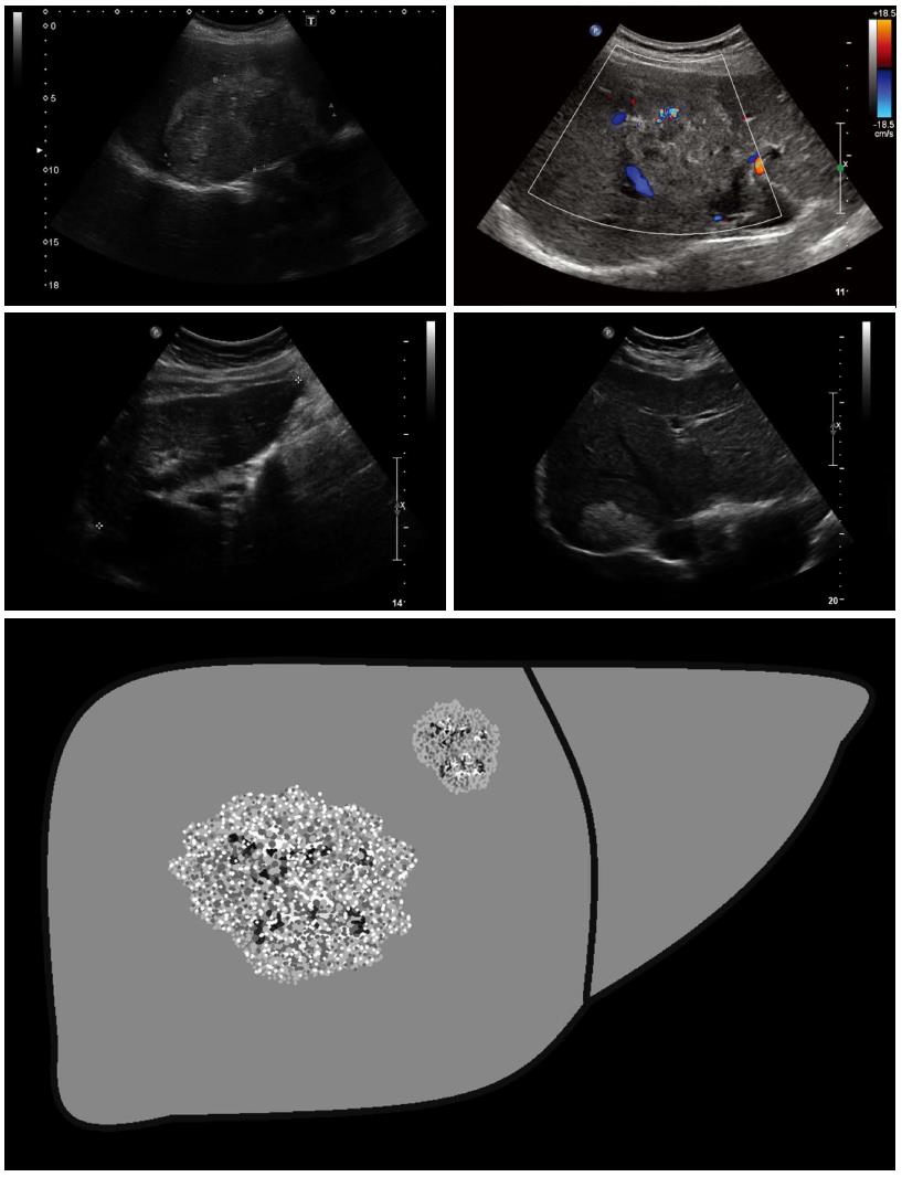
Figure 3 Hemangioma-like: These lesions are difficult to distinguish from atypical (e.
g., partially thrombosed) hemangiomas, and often represent a significant diagnostic challenge. Sonomorphologically, the lesions present as a relatively clearly demarcated non-homogeneous tumor that appears hyperechoic in comparison with the surrounding hepatic parenchyma. Echogenicity ranges from slightly and non-homogeneously hyperechoic to strongly and homogeneous hyperechoic.
- Citation: Kratzer W, Gruener B, Kaltenbach TE, Ansari-Bitzenberger S, Kern P, Fuchs M, Mason RA, Barth TF, Haenle MM, Hillenbrand A, Oeztuerk S, Graeter T. Proposal of an ultrasonographic classification for hepatic alveolar echinococcosis: Echinococcosis multilocularis Ulm classification-ultrasound. World J Gastroenterol 2015; 21(43): 12392-12402
- URL: https://www.wjgnet.com/1007-9327/full/v21/i43/12392.htm
- DOI: https://dx.doi.org/10.3748/wjg.v21.i43.12392


