Copyright
©The Author(s) 2015.
World J Gastroenterol. Oct 21, 2015; 21(39): 11053-11076
Published online Oct 21, 2015. doi: 10.3748/wjg.v21.i39.11053
Published online Oct 21, 2015. doi: 10.3748/wjg.v21.i39.11053
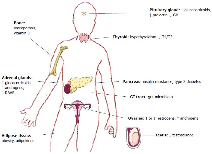
Figure 1 Endocrine diseases associated with nonalcoholic fatty liver disease.
GH: Growth hormone; RAAS: Renin-angiotensin-aldosterone system; GI: Gastrointestinal.
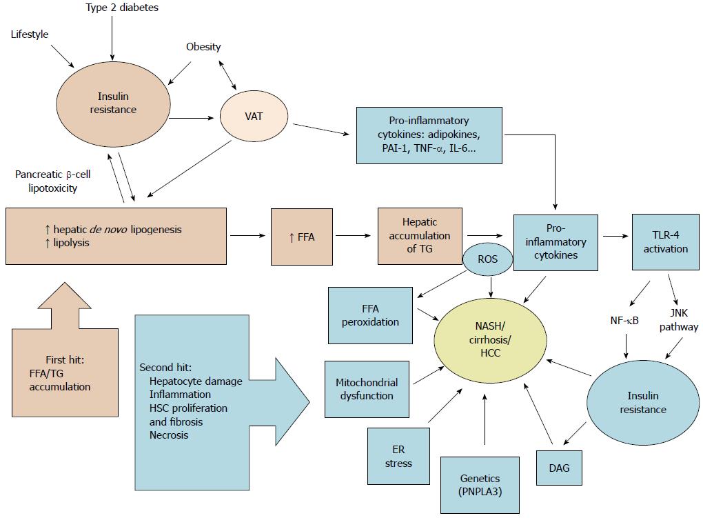
Figure 2 Schematic summary of nonalcoholic fatty liver disease pathophysiology according to the “two-hit hypothesis”.
VAT: Visceral adipose tissue; FFA: Free fatty acid; TG: Triglycerides; PAI-1: Plasminogen activator inhibitor-1; TNF-α: Tumor necrosis factor α; IL-6: Interleukin 6; ROS: Reactive oxygen species; TLR-4: Toll-like receptor 4; DAG: Diacylglycerols; ER: Endoplasmic reticulum; NASH: Non-alcoholic steatohepatitis; HCC: Hepatocellular carcinoma; PNPLA3: Patatin-like phospholipase domain-containing protein 3; NF-κB: Nuclear factor-kappa B; JNK: c-Jun N-terminal kinases; HSC: Hepatic stellate cells.
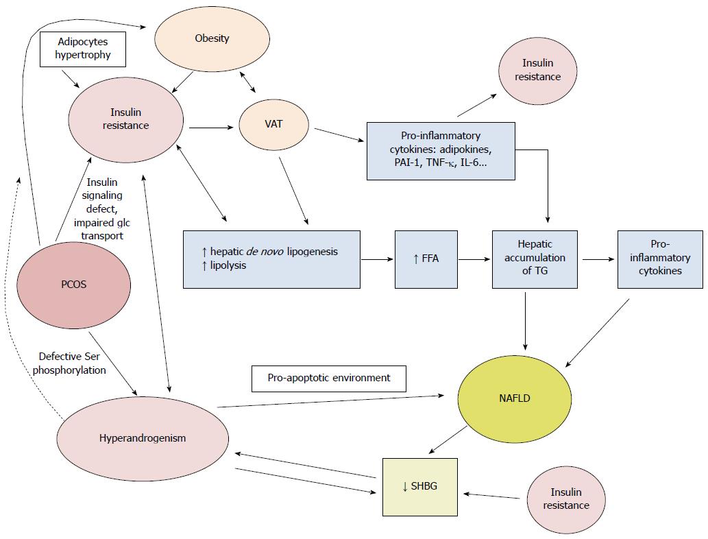
Figure 3 Pathophysiological mechanisms linking polycystic ovary syndrome and nonalcoholic fatty liver disease.
Ser: Serine; VAT: Visceral adipose tissue; glc: Glucose; SHBG: Sex hormone binding globulin; FFA: Free fatty acids; TG: Triglycerides; PAI-1: Plasminogen activator inhibitor 1; TNF-α: Tumor necrosis factor α; IL-6: Interleukin 6.
- Citation: Marino L, Jornayvaz FR. Endocrine causes of nonalcoholic fatty liver disease. World J Gastroenterol 2015; 21(39): 11053-11076
- URL: https://www.wjgnet.com/1007-9327/full/v21/i39/11053.htm
- DOI: https://dx.doi.org/10.3748/wjg.v21.i39.11053


