Copyright
©The Author(s) 2015.
World J Gastroenterol. Oct 7, 2015; 21(37): 10704-10708
Published online Oct 7, 2015. doi: 10.3748/wjg.v21.i37.10704
Published online Oct 7, 2015. doi: 10.3748/wjg.v21.i37.10704
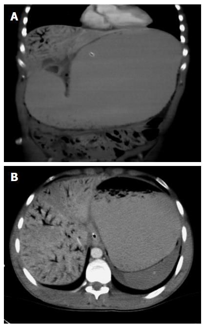
Figure 1 Abdominal computerized tomography shows massive gastric dilatation and hepatic portal venous gas at presentation.
A: Coronal section; B: Transverse section.
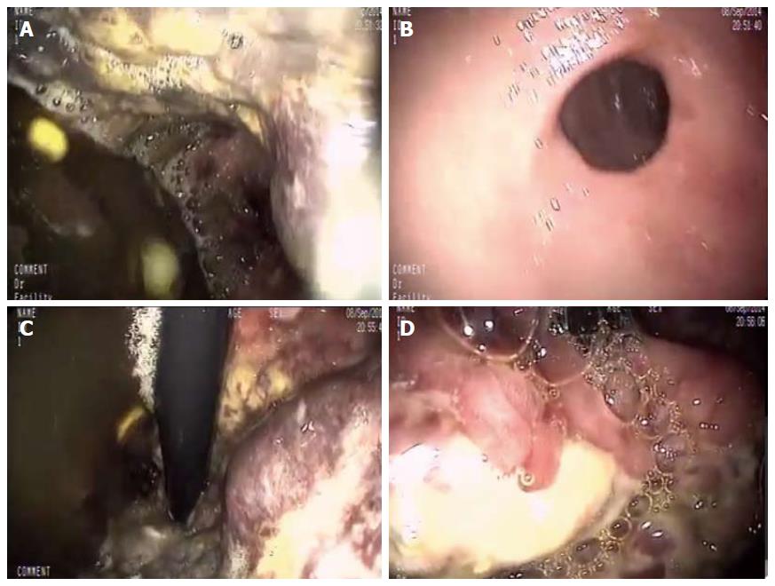
Figure 2 Patchy ischemia of gastric mucosa in emergency upper gastrointestinal endoscopy at presentation.
A: Entry to stomach; B: Pylorus; C: Corpus, view of retroflexion; D: Cardia, view of retroflexion.
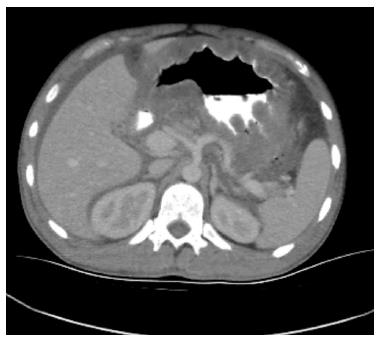
Figure 3 Transverse section of abdominal computerized tomography after 24 h.
Hepatic portal venous gas and gastric dilatation regressed dramatically.
- Citation: Sevinc MM, Kinaci E, Bayrak S, Yardimci AH, Cakar E, Bektaş H. Extraordinary cause of acute gastric dilatation and hepatic portal venous gas: Chronic use of synthetic cannabinoid. World J Gastroenterol 2015; 21(37): 10704-10708
- URL: https://www.wjgnet.com/1007-9327/full/v21/i37/10704.htm
- DOI: https://dx.doi.org/10.3748/wjg.v21.i37.10704


