Copyright
©The Author(s) 2015.
World J Gastroenterol. Jul 7, 2015; 21(25): 7929-7932
Published online Jul 7, 2015. doi: 10.3748/wjg.v21.i25.7929
Published online Jul 7, 2015. doi: 10.3748/wjg.v21.i25.7929
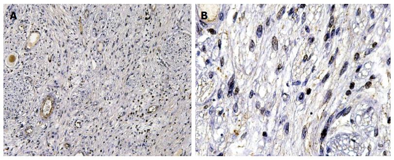
Figure 1 Immunohistochemical staining showing vasoactive intestinal peptide positivity.
A: The vasoactive intestinal peptide mainly expressed in cytoplasm [hematoxylin-eosin (HE) × 4]; B: The cytoplasm was dyed brown (HE × 100).
- Citation: Han W, Wang HM. Refractory diarrhea: A paraneoplastic syndrome of neuroblastoma. World J Gastroenterol 2015; 21(25): 7929-7932
- URL: https://www.wjgnet.com/1007-9327/full/v21/i25/7929.htm
- DOI: https://dx.doi.org/10.3748/wjg.v21.i25.7929


