Copyright
©The Author(s) 2015.
World J Gastroenterol. Jul 7, 2015; 21(25): 7907-7910
Published online Jul 7, 2015. doi: 10.3748/wjg.v21.i25.7907
Published online Jul 7, 2015. doi: 10.3748/wjg.v21.i25.7907
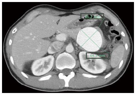
Figure 1 Abdominal computed tomography images demonstrating a splenic vein aneurysm approximately 56 mm × 64 mm in size.
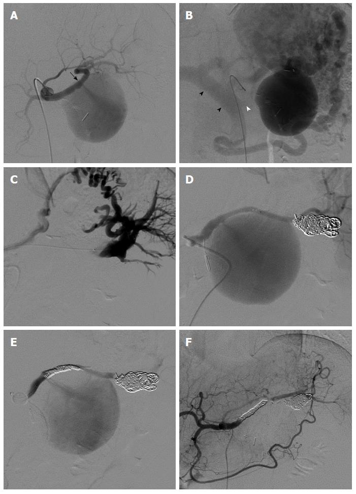
Figure 2 Angiographic imaging.
A: Celiac artery angiogram demonstrating a fistula (black arrow) between the proximal splenic artery and the splenic venous aneurysm; B: The contrast medium from the splenic vein aneurysm continued to the portal vein (black arrowhead) through the collateral vessels; the splenic vein main trunk on the portal vein side was occluded (white arrowhead); C: A micro-catheter is shown advancing to the drainage vein through the aneurysm; D: The drainage vein after embolization with 7 coils; E: The splenic artery, including the splenic arteriovenous fistula, after coil embolization; F: The splenic vein aneurysm disappeared after embolization of the splenic artery, including the splenic arteriovenous fistula and drainage vein.
- Citation: Ueda T, Murata S, Yamamoto A, Tamai J, Kobayashi Y, Hiranuma C, Yoshida H, Kumita SI. Endovascular treatment of post-laparoscopic pancreatectomy splenic arteriovenous fistula with splenic vein aneurysm. World J Gastroenterol 2015; 21(25): 7907-7910
- URL: https://www.wjgnet.com/1007-9327/full/v21/i25/7907.htm
- DOI: https://dx.doi.org/10.3748/wjg.v21.i25.7907


