Copyright
©The Author(s) 2015.
World J Gastroenterol. May 21, 2015; 21(19): 5856-5866
Published online May 21, 2015. doi: 10.3748/wjg.v21.i19.5856
Published online May 21, 2015. doi: 10.3748/wjg.v21.i19.5856
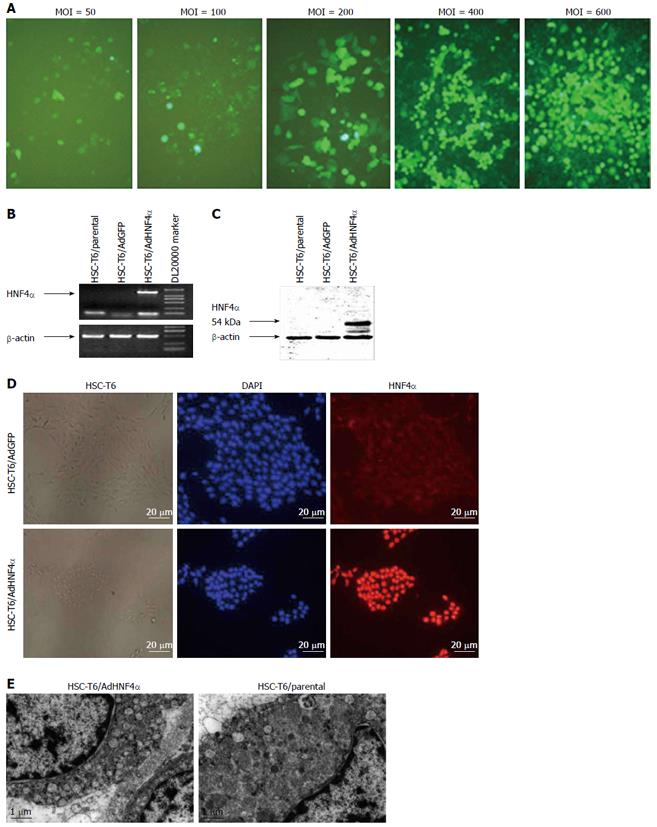
Figure 1 Ad-hepatocyte nuclear factor 4α-mediated hepatocyte nuclear factor 4α expression in rat hepatic stellate cells-T6 cells.
A: AdGFP was transfected to HSC-T6 at multiplicities of infection (MOIs) of 50, 100, 200, 400, and 600 pfu/mL. After 72 h, the GFP-positive cells were counted under a microscope. The transfection efficiency was proportional to the MOIs; original magnification × 200 ×; B-D: 72 h after transfection of AdHNF4α, HNF4α expression in HSC-T6 cells was detected by RT-PCR (B), Western blotting (C), and immunofluorescence (D); original magnification × 100; The cells were harvested and fixed in 4% paraformaldehyde and 1% glutaraldehyde, and the EPON 812-embedded ultra-thin sections were observed under transmission electron microscope (E). HNF4α: Hepatocyte nuclear factor 4α; HSCs: Hepatic stellate cells.
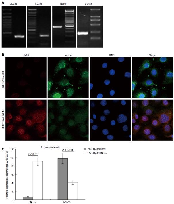
Figure 2 Identification of the stemness of rat hepatic stellate cells.
A: The expression of all the stem cell markers (CD133, CD105, and nestin) was positive in HSC-T6 cells; B: After transfection of adenovirus AdHNF4α, the expression of HNF4α and Nanog was observed by co-focal immunofluorescent staining; original magnification × 400; C: The relative expression levels of HNF4α and Nanog were calculated by image density analysis with the Image-Pro Plus V6.0 (Media Cybernetics, Inc., Rockville, MD, United States) normalized with DAPI staining. HNF4α: Hepatocyte nuclear factor 4α; HSCs: Hepatic stellate cells.
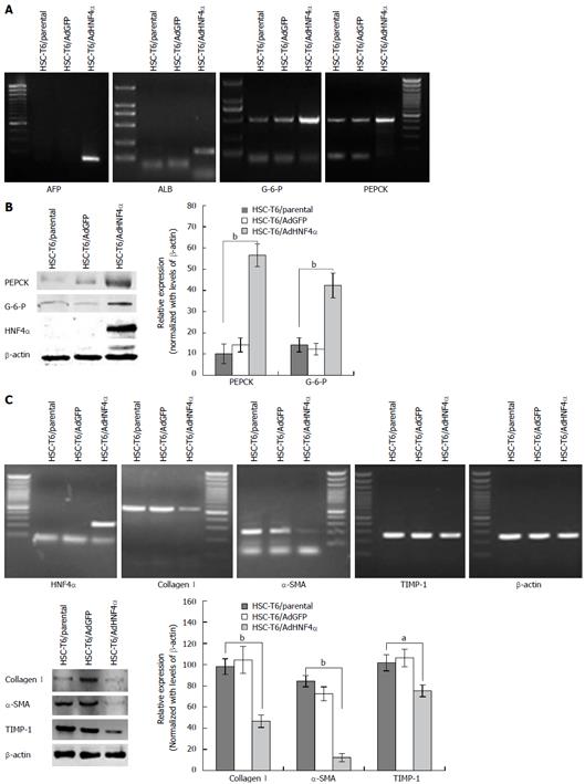
Figure 3 Identification of hepatic stellate cells differentiation mediated by hepatocyte nuclear factor 4α expression.
By reverse transcription-polymerase chain reaction and Western blotting, the expression of differentiation functional genes of hepatocytes (A and B) and genes related to fibroblast cells (C) was detected in the AdHNF4α- and AdGFP-infected groups. The relative expression levels of the indicated factors were calculated by image density analysis normalized with β-actin. aP < 0.05, bP < 0.01, HSC-T6/parental vs HSC-T6/AdHNF4α. HNF4α: Hepatocyte nuclear factor 4α; HSCs: Hepatic stellate cells.
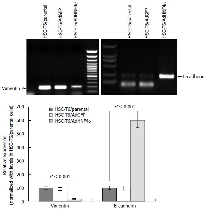
Figure 4 Hepatocyte nuclear factor 4α mediated change of epithelial-mesenchymal transition phenotypic markers in hepatic stellate cells.
After transfection of adenoviruses AdHNF4α and AdGFP, the expression of vimentin and E-cadherin was detected by RT-PCR, and relative expression was calculated by image density analysis normalized with the expression levels in HSC-T6 parental cells. HNF4α: Hepatocyte nuclear factor 4α; HSCs: Hepatic stellate cells.
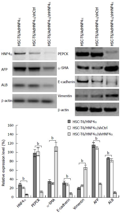
Figure 5 Effect of hepatocyte nuclear factor 4α knockout on phenotypic differentiation of hepatic stellate cells.
After HNF4α was knocked out by shHNF4α, the expression of AFP, ALB, PEPCK, E-cadherin, α-SMA, and vimentin was detected by Western blotting. β-actin was used as the control. bP < 0.01, vs control. HNF4α: Hepatocyte nuclear factor 4α.
- Citation: Liu K, Guo MG, Lou XL, Li XY, Xu Y, Ji WD, Huang XD, Yang JH, Duan JC. Hepatocyte nuclear factor 4α induces a tendency of differentiation and activation of rat hepatic stellate cells. World J Gastroenterol 2015; 21(19): 5856-5866
- URL: https://www.wjgnet.com/1007-9327/full/v21/i19/5856.htm
- DOI: https://dx.doi.org/10.3748/wjg.v21.i19.5856


