Copyright
©The Author(s) 2015.
World J Gastroenterol. Apr 14, 2015; 21(14): 4385-4390
Published online Apr 14, 2015. doi: 10.3748/wjg.v21.i14.4385
Published online Apr 14, 2015. doi: 10.3748/wjg.v21.i14.4385
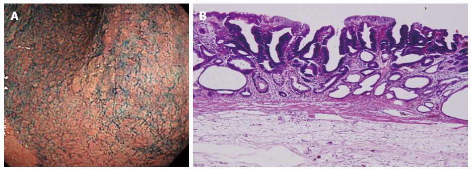
Figure 1 Initial early gastric cancer lesion in June 2005.
A: 0-IIa type well differentiated adenocarcinoma limited to the mucosa, 10 mm in size, without an ulcer scar, on the lesser curvature of the middle gastric body; B: Histopathological findings revealed a well differentiated mucosal adenocarcinoma, 10 mm in size, without lymphovascular involvement or ulcerative finding, as well as tumor-free margins.
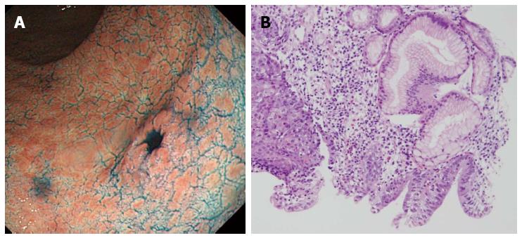
Figure 2 Metachronous early gastric cancer lesion in November 2007.
A: 0-IIc type metachronous early gastric cancer lesion, 8 mm in size, without an ulcer scar, on the greater curvature of gastric antrum. The estimated tumor depth was up to the mucosa; B: Biopsy revealed well and poorly differentiated adenocarcinoma (hematoxylin-eosin staining).
Figure 3 Histopathological findings of endoscopic submucosal dissection specimens.
A: Low magnification view with hematoxylin and eosin staining. Endoscopic submucosal dissection specimen revealed adenosquamous carcinoma invading the deep submucosal layer (1600 μm); B: High magnification view of yellow box in A. The tumor shows a solid growth pattern and prominent keratinization suggesting squamous cell carcinomatous component.
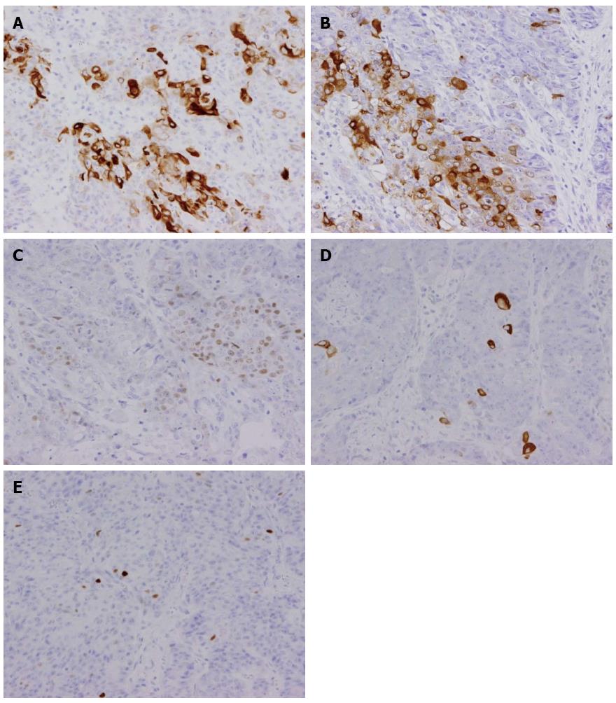
Figure 4 Immunochemical staining.
Immunohistochemical staining was focally positive for CK5/6 (A), CEA (B), CDX2 (C), and a few cells were positive for CK14 (D), P63 (E).
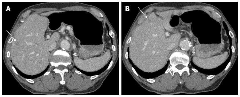
Figure 5 Computerized tomography in January 2008 (two months after the endoscopic submucosal dissection).
A and B: Enhanced computerized tomography revealed multiple low density areas suggesting liver metastases (indicated by arrows).
- Citation: Shirahige A, Suzuki H, Oda I, Sekiguchi M, Mori G, Abe S, Nonaka S, Yoshinaga S, Sekine S, Kushima R, Saito Y, Fukagawa T, Katai H. Fatal submucosal invasive gastric adenosquamous carcinoma detected at surveillance after gastric endoscopic submucosal dissection. World J Gastroenterol 2015; 21(14): 4385-4390
- URL: https://www.wjgnet.com/1007-9327/full/v21/i14/4385.htm
- DOI: https://dx.doi.org/10.3748/wjg.v21.i14.4385


