Copyright
©The Author(s) 2015.
World J Gastroenterol. Apr 14, 2015; 21(14): 4385-4390
Published online Apr 14, 2015. doi: 10.3748/wjg.v21.i14.4385
Published online Apr 14, 2015. doi: 10.3748/wjg.v21.i14.4385
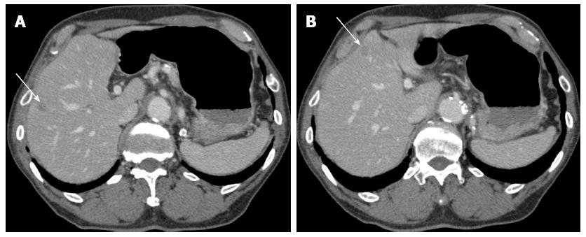
Figure 5 Computerized tomography in January 2008 (two months after the endoscopic submucosal dissection).
A and B: Enhanced computerized tomography revealed multiple low density areas suggesting liver metastases (indicated by arrows).
- Citation: Shirahige A, Suzuki H, Oda I, Sekiguchi M, Mori G, Abe S, Nonaka S, Yoshinaga S, Sekine S, Kushima R, Saito Y, Fukagawa T, Katai H. Fatal submucosal invasive gastric adenosquamous carcinoma detected at surveillance after gastric endoscopic submucosal dissection. World J Gastroenterol 2015; 21(14): 4385-4390
- URL: https://www.wjgnet.com/1007-9327/full/v21/i14/4385.htm
- DOI: https://dx.doi.org/10.3748/wjg.v21.i14.4385


