Copyright
©The Author(s) 2015.
World J Gastroenterol. Apr 14, 2015; 21(14): 4275-4283
Published online Apr 14, 2015. doi: 10.3748/wjg.v21.i14.4275
Published online Apr 14, 2015. doi: 10.3748/wjg.v21.i14.4275
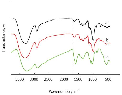
Figure 1 Fourier transform infrared spectrometer spectra of chemical groups of pure dextran (a), pure dextran coated ultrasmall superparamagnetic iron oxide nanoparticles (b), and oxidized dextran coated ultrasmall superparamagnetic iron oxide nanoparticles (c).
The peak of 1615 indicates carboxylate groups (dot line).
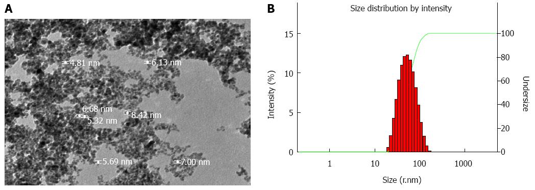
Figure 2 The properties of the magnetic molecular probes.
A: Transmission electron microscopy demonstrates the size and morphology of the magnetic molecular probes under a magnification of × 40000; B: Malvern Zetasizer Nano ZS90 laser granulometer showed the mean hydrodynamic diameter of the magnetic molecular probes and their distribution.
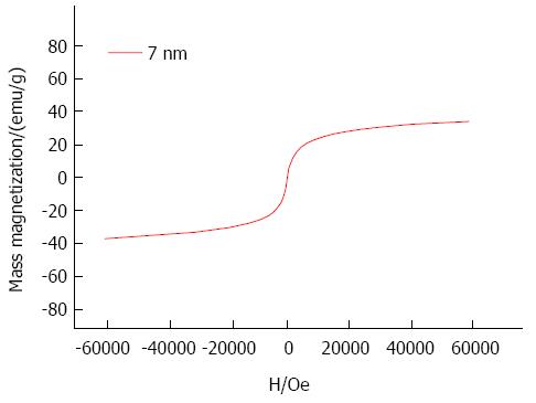
Figure 3 Hysteresis loops of the 7 nm (red line) magnetic probes at 0.
6 T.
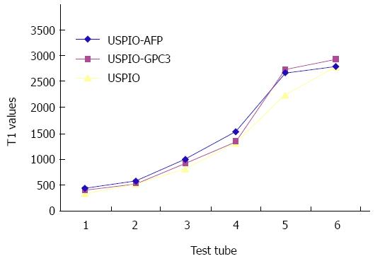
Figure 4 Relationship between T1 values and samples with different concentrations.
The test tubes 1-5 contain the iron concentrations of 0.25, 0.125, 0.0625, 0.031 and 0.016 mg/mL in the anti-AFP-USPIO, anti-GPC3-USPIO or USPIO solution, respectively. The test tube 6 contains 1% agar solution as control. USPIO: Ultrasmall superparamagnetic iron oxide; USPIO-AFP: USPIO-α-fetoprotein; USPIO-GPC3: USPIO-glypican 3.
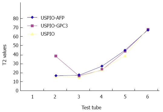
Figure 5 Relationship between T2 values and samples with different concentrations.
The test tubes 1-5 contain the iron concentrations of 0.25, 0.125, 0.0625, 0.031 and 0.016 mg/mL in the anti-AFP-USPIO, anti-GPC3-USPIO or USPIO solution, respectively. The test tube 6 contains 1% agar solution as control. USPIO: Ultrasmall superparamagnetic iron oxide; USPIO-AFP: USPIO-α-fetoprotein; USPIO-GPC3: USPIO-glypican 3.
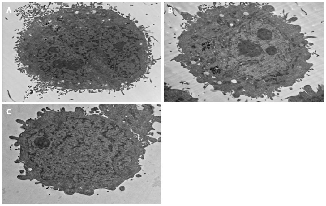
Figure 6 Transmission electron microscopy demonstrates the structure of the HepG2 cells under a magnification of × 10000.
A: HepG2 cell with expression of GPC3 receptors incubated with anti-GPC3-USPIONs probes, and the probes were seen on the surface of the cell and in the cell (black aggregation); B: HepG2 cell with expression of AFP receptors incubated with anti-AFP-USPIONs probes, and the probes were seen in the cell (black aggregation); C: HepG2 cell with expression of GPC3 receptors incubated with USPIONs probes, and no USPION was seen. USPION: Ultrasmall superparamagnetic iron oxide nanoparticle; AFP: α-fetoprotein; GPC3: Glypican 3.
- Citation: Li YW, Chen ZG, Zhao ZS, Li HL, Wang JC, Zhang ZM. Preparation of magnetic resonance probes using one-pot method for detection of hepatocellular carcinoma. World J Gastroenterol 2015; 21(14): 4275-4283
- URL: https://www.wjgnet.com/1007-9327/full/v21/i14/4275.htm
- DOI: https://dx.doi.org/10.3748/wjg.v21.i14.4275


