Copyright
©The Author(s) 2015.
World J Gastroenterol. Mar 28, 2015; 21(12): 3755-3758
Published online Mar 28, 2015. doi: 10.3748/wjg.v21.i12.3755
Published online Mar 28, 2015. doi: 10.3748/wjg.v21.i12.3755
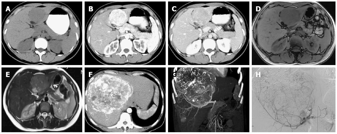
Figure 1 Imaging findings.
A: The tumor was hypoechoic on computed tomography (CT); B, C: Enhanced CT showed a tumor (7.0 cm × 9.0 cm) with early-phase hyperattenuation and late-phase hypoattenuation; D, E: Magnetic resonance imaging showed a tumor with hypointensity on T1-weighted, hyperintensity on T2-weighted; F, G: Enhanced CT showed two hepatic masses (13.0 cm × 12.0 cm and 2.3 cm × 1.8 cm) in the right lobe; H: Angiography revealed a circumscribed hypervascular mass.
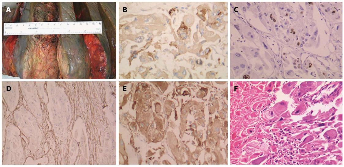
Figure 2 Pathologic findings.
A: The tumor occupied a large area of the liver; The tumor was immunopositive for B: Human melanoma black-45 (20 ×); C: Ki-67 (10 ×); D: Smooth muscle actin (10 ×); E: CD68 (20 ×); F: Hematoxylin and eosin staining showed neoplasm necrosis (4 ×).
- Citation: Wang WT, Li ZQ, Zhang GH, Guo Y, Teng MJ. Liver transplantation for recurrent posthepatectomy malignant hepatic angiomyolipoma: A case report. World J Gastroenterol 2015; 21(12): 3755-3758
- URL: https://www.wjgnet.com/1007-9327/full/v21/i12/3755.htm
- DOI: https://dx.doi.org/10.3748/wjg.v21.i12.3755


