Copyright
©The Author(s) 2015.
World J Gastroenterol. Mar 28, 2015; 21(12): 3607-3613
Published online Mar 28, 2015. doi: 10.3748/wjg.v21.i12.3607
Published online Mar 28, 2015. doi: 10.3748/wjg.v21.i12.3607
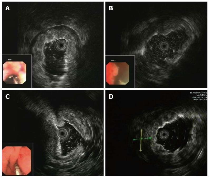
Figure 1 Endoscopic ultrasound characteristics of gastric infiltrating tumors.
A: Invaded muscularis propria and the first three blurred sonographic layers; B: Invaded serosal layer; C: Ascites around gastric wall; D: Perigastric lymph node.
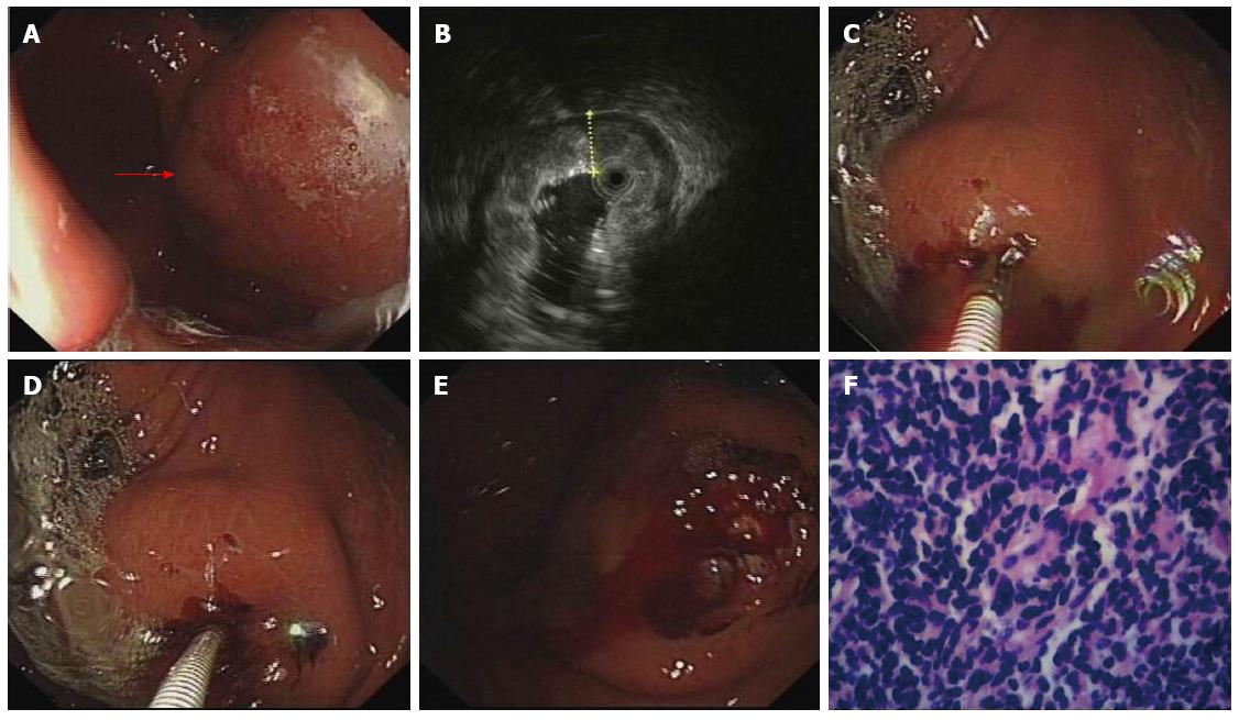
Figure 2 Endoscopic ultrasound-guided bite-on-bite technique.
A and B: A gastric infiltrating tumor diagnosed by endoscopic ultrasound (EUS) (red arrow); C: Bite-on-bite technique performed after EUS localization; D: The bite is taken on top of the previous one to obtain valid tissue; E: Postoperative wound; F: Histology confirmed lymphoma (HE staining, × 400).
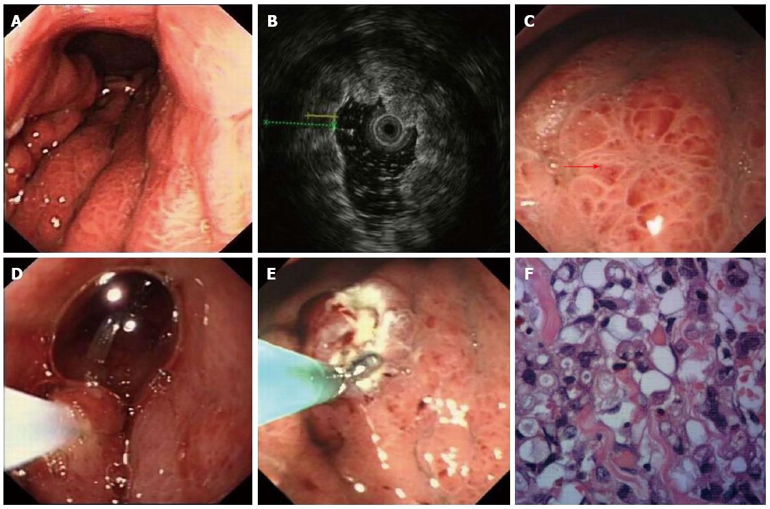
Figure 3 Endoscopic mucosal resection combined with bite-on-bite technique under the guidance of endoscopic ultrasound.
A and B: A gastric infiltrating tumor diagnosed by endoscopic ultrasound (EUS); C: The thickest site was selected for biopsy after EUS localization (red arrow); D: The mucosa was then resected; E: Bite-on-bite technique was performed in the resection area; F: Histology confirmed adenocarcinoma with poor differentiation (HE staining × 400).
- Citation: Zhou XX, Pan HH, Usman A, Ji F, Jin X, Zhong WX, Chen HT. Endoscopic ultrasound-guided deep and large biopsy for diagnosis of gastric infiltrating tumors with negative malignant endoscopy biopsies. World J Gastroenterol 2015; 21(12): 3607-3613
- URL: https://www.wjgnet.com/1007-9327/full/v21/i12/3607.htm
- DOI: https://dx.doi.org/10.3748/wjg.v21.i12.3607


