Copyright
©The Author(s) 2015.
World J Gastroenterol. Mar 21, 2015; 21(11): 3435-3440
Published online Mar 21, 2015. doi: 10.3748/wjg.v21.i11.3435
Published online Mar 21, 2015. doi: 10.3748/wjg.v21.i11.3435
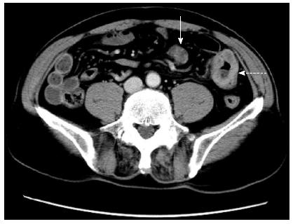
Figure 1 Abdominal computed tomography scan revealed metastatic tumor mass of ileum (solid arrow) and the enlarged lymph node (dotted arrow).
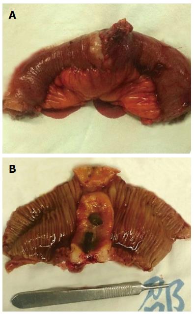
Figure 2 Intraoperative imaging of the resected tumor of the ileum.
The tumor was 4.5 cm × 3.0 cm in size, with a clear margin and ulceration on the intraluminal surface.
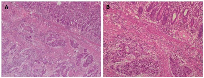
Figure 3 Microscopic findings of metastatic lung squamous cell carcinoma in the ileum.
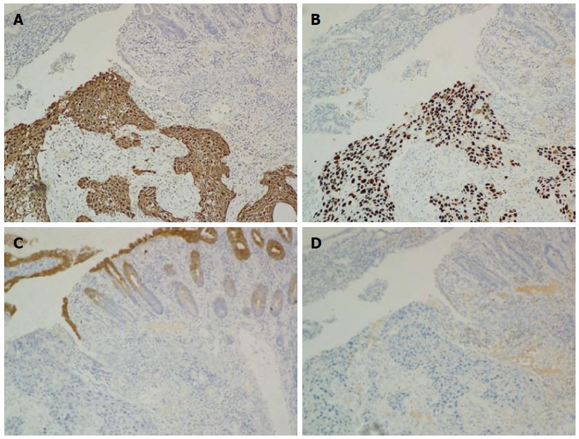
Figure 4 By immunohistochemistry, the tumor cells were found to be positive for CKH (A) and P63 (B), but negative for CK20 (C) and CK7 (D).
- Citation: Liu W, Zhou W, Qi WL, Ma YD, Xu YY. Gastrointestinal hemorrhage due to ileal metastasis from primary lung cancer. World J Gastroenterol 2015; 21(11): 3435-3440
- URL: https://www.wjgnet.com/1007-9327/full/v21/i11/3435.htm
- DOI: https://dx.doi.org/10.3748/wjg.v21.i11.3435


