Copyright
©2014 Baishideng Publishing Group Inc.
World J Gastroenterol. Dec 21, 2014; 20(47): 18048-18052
Published online Dec 21, 2014. doi: 10.3748/wjg.v20.i47.18048
Published online Dec 21, 2014. doi: 10.3748/wjg.v20.i47.18048
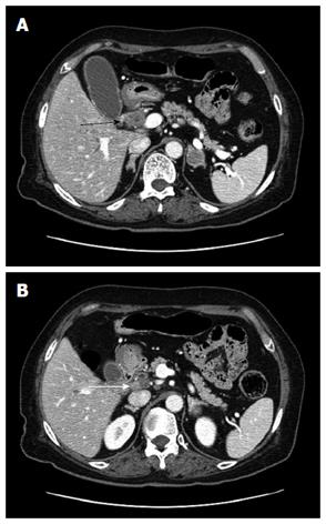
Figure 1 Computed tomography findings.
A: One mass 2.7 cm in size in the mid common bile duct (black arrow); B: Regional metastatic node in the hepatoduodenal ligament (white arrow).
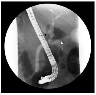
Figure 2 Endoscopic retrograde cholangiopancreatography.
The image presents an asymmetric filling defect in the middle common bile duct with proximal dilatation (black arrow) and the cystic duct was well visualized (white arrow).
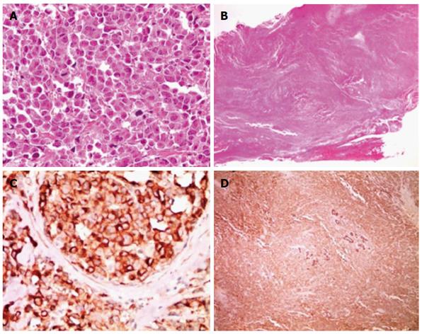
Figure 3 Histologic and immunohistochemical findings.
A: High cellularity tumor cells invades into the bile duct wall (magnification × 12.5); B: tumor cells exhibit relatively abundant cytoplasm and hyperchromatic round nuclei with course chromatin and prominent nucleoli (magnification × 400); C: tumor cells exhibit immunopositivity for a neuroendocrine marker (synaptophysin) (magnification: × 400); D: immunohistochemical staining for Pan CK, and a combined adenocarcinoma component is not noted (magnification: × 40).
- Citation: Park SB, Moon SB, Ryu YJ, Hong J, Kim YH, Chae GB, Hong SK. Primary large cell neuroendocrine carcinoma in the common bile duct: First Asian case report. World J Gastroenterol 2014; 20(47): 18048-18052
- URL: https://www.wjgnet.com/1007-9327/full/v20/i47/18048.htm
- DOI: https://dx.doi.org/10.3748/wjg.v20.i47.18048


