Copyright
©2014 Baishideng Publishing Group Inc.
World J Gastroenterol. Dec 21, 2014; 20(47): 18048-18052
Published online Dec 21, 2014. doi: 10.3748/wjg.v20.i47.18048
Published online Dec 21, 2014. doi: 10.3748/wjg.v20.i47.18048
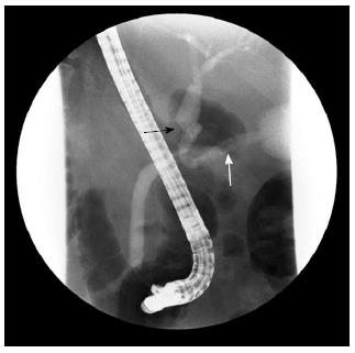
Figure 2 Endoscopic retrograde cholangiopancreatography.
The image presents an asymmetric filling defect in the middle common bile duct with proximal dilatation (black arrow) and the cystic duct was well visualized (white arrow).
- Citation: Park SB, Moon SB, Ryu YJ, Hong J, Kim YH, Chae GB, Hong SK. Primary large cell neuroendocrine carcinoma in the common bile duct: First Asian case report. World J Gastroenterol 2014; 20(47): 18048-18052
- URL: https://www.wjgnet.com/1007-9327/full/v20/i47/18048.htm
- DOI: https://dx.doi.org/10.3748/wjg.v20.i47.18048


