Copyright
©2014 Baishideng Publishing Group Inc.
World J Gastroenterol. Dec 21, 2014; 20(47): 17727-17736
Published online Dec 21, 2014. doi: 10.3748/wjg.v20.i47.17727
Published online Dec 21, 2014. doi: 10.3748/wjg.v20.i47.17727
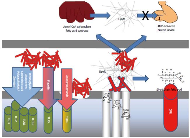
Figure 2 Examples of some theories on potential pathways for the impact of gut microbiota on animal models of human disease.
Bacterial colonization may double the density of capillaries in the small intestinal epithelium, thereby promoting intestinal monosaccharide absorption[28]. Undigested food components may be fermented into SCFAs and subsequently act as signals for GPRs of importance for the development of obesity[26,29]. Bacteria may express several key enzymes relevant for hepatic lipogenesis[27,50], and hepatic and muscular fatty acid oxidation[31]. Molecular structures in the cell walls of bacteria may act as MAMPs, which stimulate TLRs on the host cells to induce innate immune responses. The complex of TLR1, TLR2, TLR6 and TLR10 is expressed on a range of cell types such as enterocytes, macrophages, dendritic cells, natural killer cells, mast cells, T cells, B cells, neutrophilic cells and Schwann cells, and may be stimulated by various MAMPs, e.g. peptidoglycan, from Gram positive bacteria cell types[21,23-25,31-33]. TLR4, expressed by e.g. macrophages, dendritic cells, mast cells, natural killer cells and enterocytes, is stimulated by lipopolysaccharides from Gram negative bacteria[30], while flagellin from various bacteria may stimulate TLR5 expressed by e.g. mucosal dendritic cells and macrophages[33]. Mucin-degrading Akkermansia muciniphila may reduce the mucus layer to increase TLR-stimulation[79]. SCFAs: Short chain fatty acids; GPR: G-protein receptor; MAMP: Microbial-associated molecular pattern; TLR: Toll-like receptor.
- Citation: Hansen AK, Hansen CHF, Krych L, Nielsen DS. Impact of the gut microbiota on rodent models of human disease. World J Gastroenterol 2014; 20(47): 17727-17736
- URL: https://www.wjgnet.com/1007-9327/full/v20/i47/17727.htm
- DOI: https://dx.doi.org/10.3748/wjg.v20.i47.17727


