Copyright
©2014 Baishideng Publishing Group Inc.
World J Gastroenterol. Dec 14, 2014; 20(46): 17661-17665
Published online Dec 14, 2014. doi: 10.3748/wjg.v20.i46.17661
Published online Dec 14, 2014. doi: 10.3748/wjg.v20.i46.17661
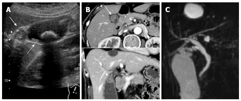
Figure 1 Diagnostic imaging results.
A: Abdominal ultrasound showing an isoechoic mass in the neck of the gallbladder and multiple small polyps measuring 3-16 mm in diameter (arrows) throughout the gallbladder (a 22 mm gallbladder stone was also identified); B: CT showed a contrast-enhanced mass (arrows) in the neck of the gallbladder; C: Magnetic resonance cholangiopancreatography showed no other abnormalities of the biliary and pancreatic ductal systems.
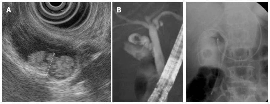
Figure 2 Endoscopic ultrasound images.
A: Endoscopic ultrasound revealed a hyperechoic papillary mass, measuring 16 mm × 8 mm in diameter, in the neck of the gallbladder, as well as multiple small polyps; B: Endoscopic naso-gallbladder drainage was performed to enable cytological examination of the bile within the gallbladder.
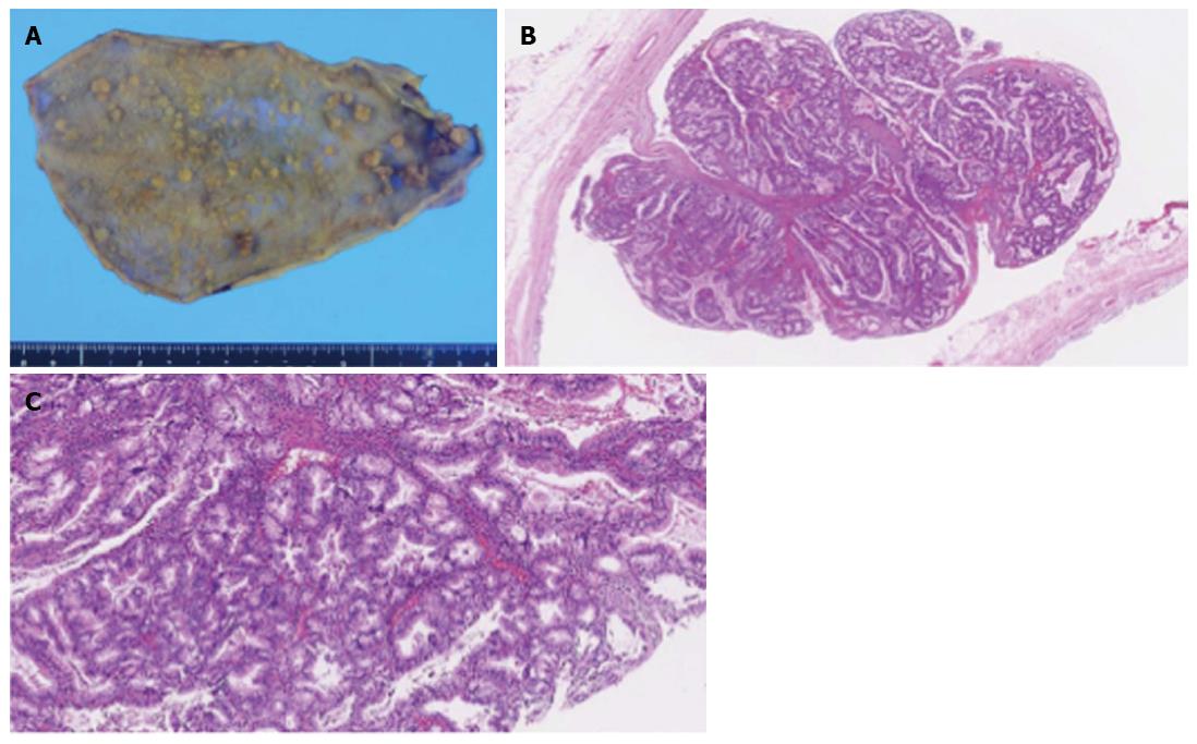
Figure 3 Pathologic examination.
A: The resected specimen showed more than 70 polypoid lesions of various sizes that were composed of proliferative lobules of closely packed pyloric-type glands displaying low- to intermediate-grade dysplasia without stromal invasion, consistent with multiple tubular adenomas; B: Hematoxylin and eosin staining (10× magnification); C: Hematoxylin and eosin staining (50× magnification).
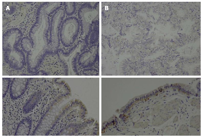
Figure 4 Immunohistochemical images.
Immunohistochemical analysis of adenomatous polyposis coli protein expression in A: Colonic polyps (upper panel, resected 22 years previously) and B: gallbladder polyps (upper panel) revealed loss of expression in both lesions compared with the normal mucosa in the colon (A: lower panel) and gallbladder (B: lower panel), which were weakly positive for adenomatous polyposis coli.
- Citation: Mori Y, Sato N, Matayoshi N, Tamura T, Minagawa N, Shibao K, Higure A, Nakamoto M, Taguchi M, Yamaguchi K. Rare combination of familial adenomatous polyposis and gallbladder polyps. World J Gastroenterol 2014; 20(46): 17661-17665
- URL: https://www.wjgnet.com/1007-9327/full/v20/i46/17661.htm
- DOI: https://dx.doi.org/10.3748/wjg.v20.i46.17661


