Copyright
©2014 Baishideng Publishing Group Inc.
World J Gastroenterol. Oct 28, 2014; 20(40): 14589-14597
Published online Oct 28, 2014. doi: 10.3748/wjg.v20.i40.14589
Published online Oct 28, 2014. doi: 10.3748/wjg.v20.i40.14589
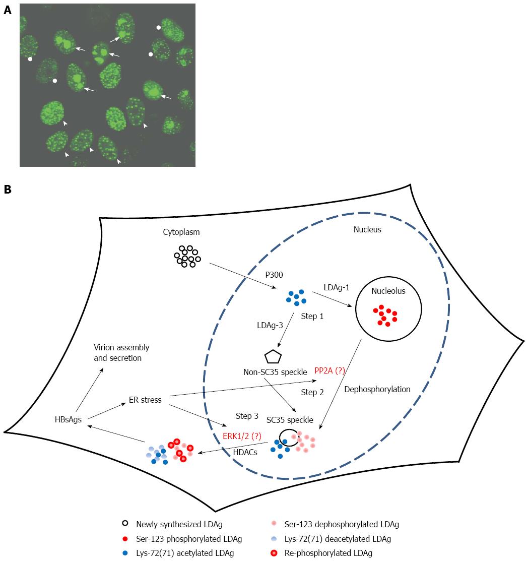
Figure 1 Large delta antigen resulting in different targeting patterns to various subcellular sites.
A: Green-fluorescent protein (GFP) fused with LDAg (GFP-LDAg) distribution shown by a fluorescence microscopic image. HeLa cells were transfected with GFP-LDAg expressing construct and the GFP signal was photographed under a fluorescence microscope. The distribution of GFP-LDAg is in different nuclear compartments indicated by the GFP signal. Three predominant locations of GFP-LDAg in GFP positive cells are nucleolus (Nu), nuclear speckles (SC35) and non-SC35 nuclear speckles (NS). The cells marked with arrow, arrowhead and circle indicate the most GFP signals in Nu, SC35 and NS, respectively; B: A schematic model of HBsAgs-induced LDAg nuclear exportation. The newly synthesized LDAg is acetylated by p300 (a type of acetyltransferase) followed by targeting to the nucleolus (genotype 1) or non-SC35 speckles (genotype 3). It is unclear whether there is a direct association between acetylation and/or serine-123 phosphorylation and LDAg localization to the nucleolus or nuclear speckles. Nevertheless, in both hepatitis D virus genotypes, LDAg moves to the SC35 speckles later (step 2) and it is speculated that phosphatase (PP2A) might contribute to the dephosphorylation of LDAgs. Afterwards, LDAg is deacetylated by histone deacetylases (HDACs) and exported out of the nucleus (step 3) and the ERK1/2 might participate in the re-phosphorylation of LDAgs. Finally, in the cytoplasm, the LDAg interacts with hepatitis B virus supplying surface antigens (HBsAgs) to assemble into empty viral particles or virions.
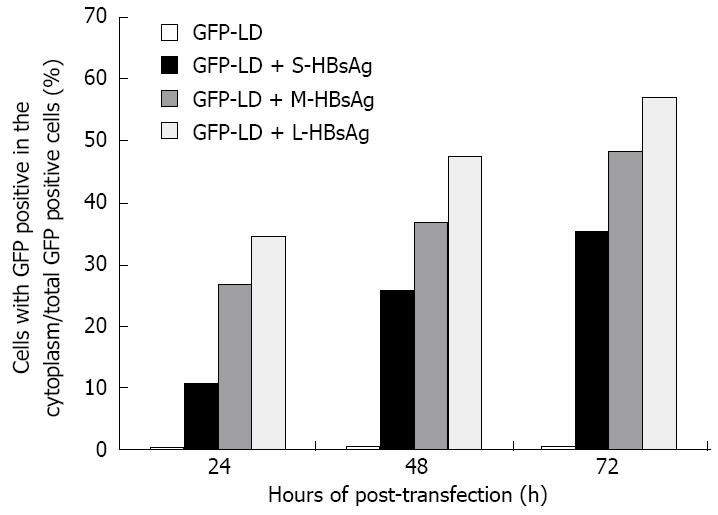
Figure 2 Distribution ratio of cytoplasmic green-fluorescent protein-large delta antigen in the presence of hepatitis B virus supplying surface antigens (modified from the results in reference 84).
The green-fluorescent protein fused with large delta antigen (GFP-LDAg) (genotype 1) expressing construct was co-transfected alone or with various hepatitis B virus supplying surface antigens (HBsAgs) expressing plasmids into HuH-7, a human hepatoma cell line. The S-HBsAg, M-HBsAg, and L-HBsAg represent the small HBsAg, middle HBsAg and large HBsAg, respectively. The distribution of GFP-LDAg was observed with a fluorescence microscope at different post-transfection times. The percentage of GFP-LD distributed in the cytoplasm is shown. Results conclude that the GFP-LD remains inside the nucleus in the absence of HBsAg. In contrast, in the presence of various forms of HBsAg, the amount of GFP-LD translocation from the nucleus to cytoplasm is time-dependent.
- Citation: Huang CR, Lo SJ. Hepatitis D virus infection, replication and cross-talk with the hepatitis B virus. World J Gastroenterol 2014; 20(40): 14589-14597
- URL: https://www.wjgnet.com/1007-9327/full/v20/i40/14589.htm
- DOI: https://dx.doi.org/10.3748/wjg.v20.i40.14589


