Copyright
©2014 Baishideng Publishing Group Inc.
World J Gastroenterol. Oct 28, 2014; 20(40): 14589-14597
Published online Oct 28, 2014. doi: 10.3748/wjg.v20.i40.14589
Published online Oct 28, 2014. doi: 10.3748/wjg.v20.i40.14589
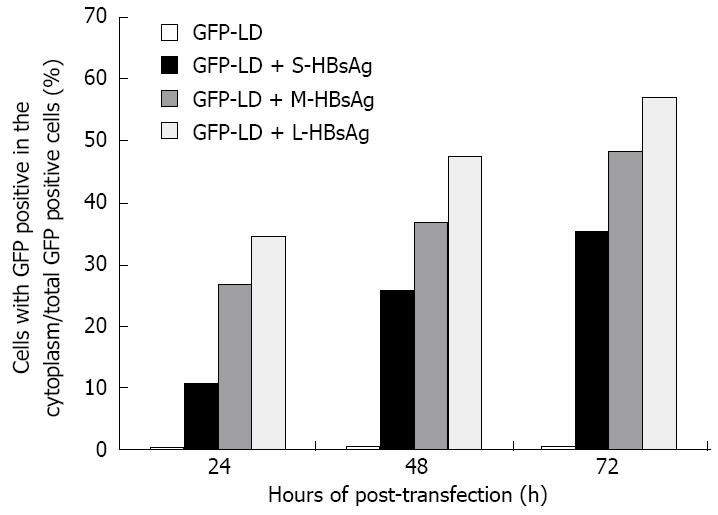
Figure 2 Distribution ratio of cytoplasmic green-fluorescent protein-large delta antigen in the presence of hepatitis B virus supplying surface antigens (modified from the results in reference 84).
The green-fluorescent protein fused with large delta antigen (GFP-LDAg) (genotype 1) expressing construct was co-transfected alone or with various hepatitis B virus supplying surface antigens (HBsAgs) expressing plasmids into HuH-7, a human hepatoma cell line. The S-HBsAg, M-HBsAg, and L-HBsAg represent the small HBsAg, middle HBsAg and large HBsAg, respectively. The distribution of GFP-LDAg was observed with a fluorescence microscope at different post-transfection times. The percentage of GFP-LD distributed in the cytoplasm is shown. Results conclude that the GFP-LD remains inside the nucleus in the absence of HBsAg. In contrast, in the presence of various forms of HBsAg, the amount of GFP-LD translocation from the nucleus to cytoplasm is time-dependent.
- Citation: Huang CR, Lo SJ. Hepatitis D virus infection, replication and cross-talk with the hepatitis B virus. World J Gastroenterol 2014; 20(40): 14589-14597
- URL: https://www.wjgnet.com/1007-9327/full/v20/i40/14589.htm
- DOI: https://dx.doi.org/10.3748/wjg.v20.i40.14589


