Copyright
©2014 Baishideng Publishing Group Inc.
World J Gastroenterol. Oct 14, 2014; 20(38): 14068-14072
Published online Oct 14, 2014. doi: 10.3748/wjg.v20.i38.14068
Published online Oct 14, 2014. doi: 10.3748/wjg.v20.i38.14068
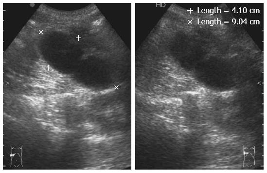
Figure 1 Abdominal ultrasonography.
Ultrasonography revealed a large, thick-walled gallbladder and the absence of gallstones.
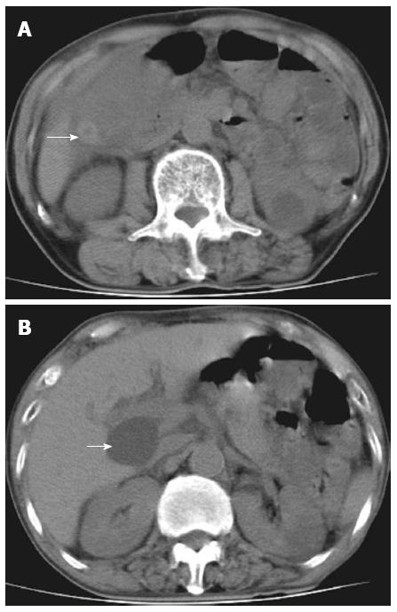
Figure 2 Abdominal computed tomography scan.
A: Computed tomography scan of abdomen in axial section indicating a hyperdense lesion (arrow); B: The hepatic cyst was located in the hilar region (arrow).
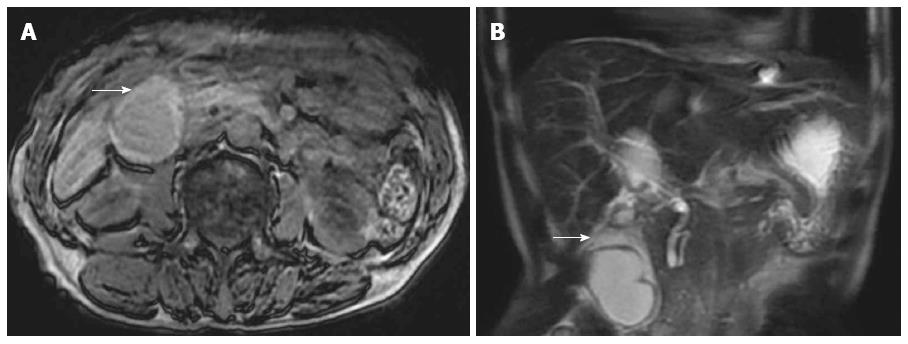
Figure 3 Abdominal T1-weighted and coronal reformatted abdominal T2-weighted magnetic resonance imaging.
A: Thickening of the gallbladder wall can be seen (arrow); B: An intra-gallbladder segment was located between the body and neck of the gallbladder (arrow), and within this segment, there was a notable crease.
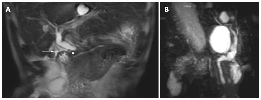
Figure 4 Magnetic resonance cholangiopancreatography results and image.
A: V-shaped distortion of the extrahepatic bile ducts (arrowhead) and tapered cystic duct (arrow) were observed; B: Choledochal cyst was excluded.
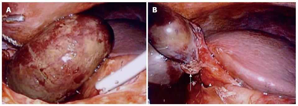
Figure 5 Laparoscopy.
A: Gallbladder appeared gangrenous; B: Gallbladder was not totally attached to the liver bed and had twisted approximately 180° in the clockwise direction (arrow). Hartmann’s pouch and the gallbladder neck were attached to the inferior surface of the liver (arrowhead).
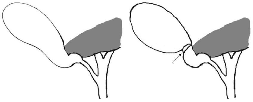
Figure 6 Gross type 1 floating gallbladder.
Clockwise torsion was observed at the neck-body junction of the gallbladder (arrow), as the result of the gallbladder being attached to the inferior surface of the liver via the mesentery. This condition allowed the gallbladder to hang free in an unstable state.
- Citation: Pu TW, Fu CY, Lu HE, Cheng WT. Complete body-neck torsion of the gallbladder: A case report. World J Gastroenterol 2014; 20(38): 14068-14072
- URL: https://www.wjgnet.com/1007-9327/full/v20/i38/14068.htm
- DOI: https://dx.doi.org/10.3748/wjg.v20.i38.14068


