
















©2014 Baishideng Publishing Group Inc.
World J Gastroenterol. Oct 14, 2014; 20(38): 14068-14072
Published online Oct 14, 2014. doi: 10.3748/wjg.v20.i38.14068
Published online Oct 14, 2014. doi: 10.3748/wjg.v20.i38.14068
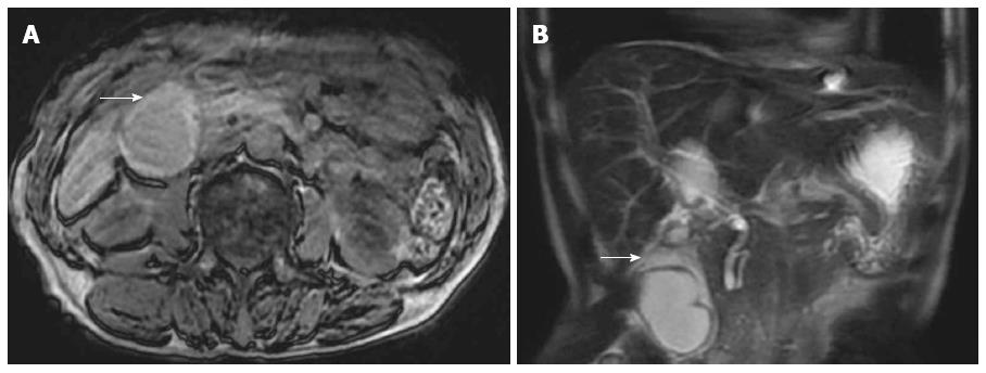
Figure 3 Abdominal T1-weighted and coronal reformatted abdominal T2-weighted magnetic resonance imaging.
A: Thickening of the gallbladder wall can be seen (arrow); B: An intra-gallbladder segment was located between the body and neck of the gallbladder (arrow), and within this segment, there was a notable crease.
- Citation: Pu TW, Fu CY, Lu HE, Cheng WT. Complete body-neck torsion of the gallbladder: A case report. World J Gastroenterol 2014; 20(38): 14068-14072
- URL: https://www.wjgnet.com/1007-9327/full/v20/i38/14068.htm
- DOI: https://dx.doi.org/10.3748/wjg.v20.i38.14068