Copyright
©2014 Baishideng Publishing Group Inc.
World J Gastroenterol. Sep 28, 2014; 20(36): 13195-13199
Published online Sep 28, 2014. doi: 10.3748/wjg.v20.i36.13195
Published online Sep 28, 2014. doi: 10.3748/wjg.v20.i36.13195
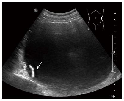
Figure 1 Abdominal ultrasonography shows a cystic mass with luminal calcification (white arrow) that measures 14 cm in diameter in the left retroperitoneum.
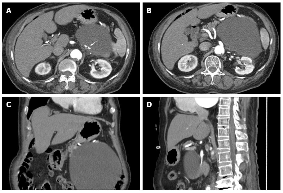
Figure 2 Computed tomography scans show a large low-attenuation lesion measuring 16 cm in the pancreatic tail with luminal calcification (white arrow) (A), the cyst abutting the pancreas (B), a large cystic mass in the retroperitoneum (C); the cystic mass below the pancreas (D).
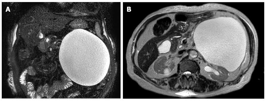
Figure 3 Magnetic resonance imaging images reveal a large cystic lesion on T2-weighted images (A), and the cyst could be observed abutting the pancreas (B).
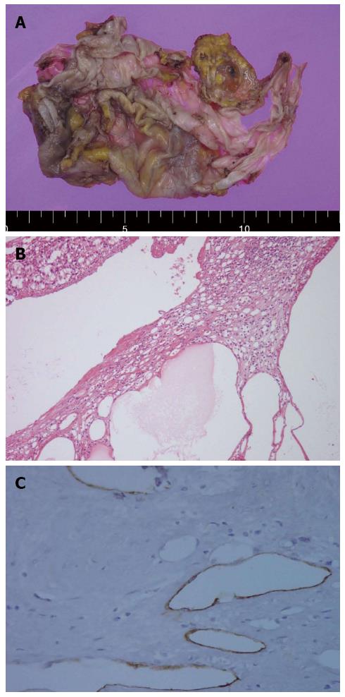
Figure 4 Pathologic pictures.
A: The gross size of the cyst was 13.0 cm × 8.0 cm, and the adrenal gland contained smaller cysts with a thin membranous appearance; B: Multiple thin-walled cysts of variable size, with remaining adrenal tissue in the intervening septae of the cysts (hematoxylin-eosin staining, × 100); C: Immunostaining with the D2-40 antibody is positive along the lining of the cysts, thus revealing lymphatic vessels.
- Citation: Jung HI, Ahn T, Son MW, Kim Z, Bae SH, Lee MS, Kim CH, Cho HD. Adrenal lymphangioma masquerading as a pancreatic tail cyst. World J Gastroenterol 2014; 20(36): 13195-13199
- URL: https://www.wjgnet.com/1007-9327/full/v20/i36/13195.htm
- DOI: https://dx.doi.org/10.3748/wjg.v20.i36.13195


