Copyright
©2014 Baishideng Publishing Group Inc.
World J Gastroenterol. Sep 21, 2014; 20(35): 12509-12516
Published online Sep 21, 2014. doi: 10.3748/wjg.v20.i35.12509
Published online Sep 21, 2014. doi: 10.3748/wjg.v20.i35.12509
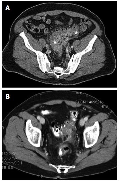
Figure 1 Computed tomography imaging demonstrating acute diverticulitis.
A: Computed tomography (CT) imaging demonstrating acute, uncomplicated diverticulitis. Reproduced with permission from Stocchi L. Current indications and role of surgery in the management of sigmoid diverticulitis. World J Gastroenterol 2010; 16(7): 804-817; B: CT imaging demonstrating acute diverticulitis with localized perforation with a small amount of extraluminal air. Reproduced with permission from Stocchi L. Current indications and role of surgery in the management of sigmoid diverticulitis. World J Gastroenterol 2010; 16 (7): 804-817.
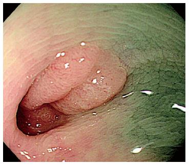
Figure 2 Colonoscopy demonstrating a polyp arising within a diverticulum in the descending colon.
Histology confirmed this to be a tubulovillous adenoma with well-differentiated adenocarcinoma confined to the mucosa. Reproduced with permission from Fu KI, Hamahata Y, Tsujinaka Y. Early colon cancer within a diverticulum treated by magnifying chromoendoscopy and laparoscopy. World J Gastroenterol 2010; 16 (12): 1545-1547.
Figure 3 Endoscopic image.
A: An inflamed diverticulum with erythema and fibrinous deposit at its orifice; B: A malignant-appearing tumor in the colon. Reproduced with permission from http://www.endoatlas.org, World Endoscopy Organization.
- Citation: Agarwal AK, Karanjawala BE, Maykel JA, Johnson EK, Steele SR. Routine colonic endoscopic evaluation following resolution of acute diverticulitis: Is it necessary? World J Gastroenterol 2014; 20(35): 12509-12516
- URL: https://www.wjgnet.com/1007-9327/full/v20/i35/12509.htm
- DOI: https://dx.doi.org/10.3748/wjg.v20.i35.12509


