Copyright
©2014 Baishideng Publishing Group Inc.
World J Gastroenterol. Sep 21, 2014; 20(35): 12473-12484
Published online Sep 21, 2014. doi: 10.3748/wjg.v20.i35.12473
Published online Sep 21, 2014. doi: 10.3748/wjg.v20.i35.12473
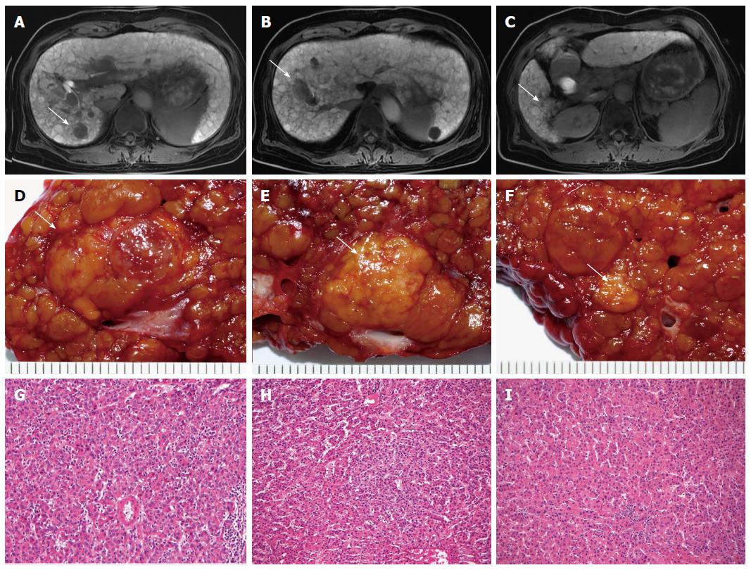
Figure 1 Sixty-nine-year-old female with cirrhosis who underwent liver resection for hepatocellular carcinoma.
A: Gd-EOB-DTPA-enhanced axial MR imaging show hypodense lesions in hepatobiliary phase in segment 7 (arrow) (tumor 1); B: Segment 8 (arrow) (tumor 2); C: Segment 6 (arrow) (tumor 3); D: Pathologic specimens of tumor 1 (arrow); E: Tumor 2 (arrow); F: Tumor 3 (arrow); G: Histology examination confirmed the diagnosis of Edmondson-Steiner grade II hepatocellular carcinoma (HCC) in tumor 1; H: Tumor 2; I: Grade I HCC in tumor 3 (HE stain, x 100).
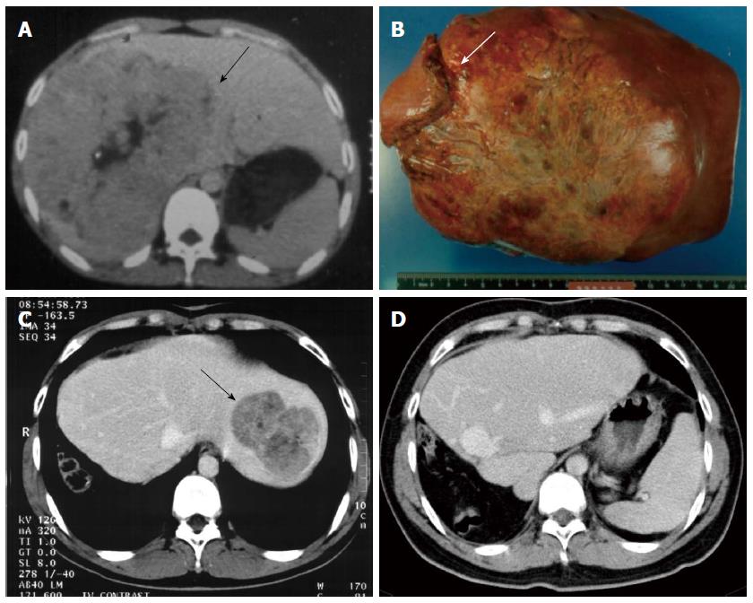
Figure 2 Eighteen-year-old male with hepatitis B virus-related hepatocellular carcinoma.
A: Computed tomography shows a 15 cm size tumor occupying nearly the entire right lobe and extending into the segment IV (arrow); B: Liver extended right lobectomy was done with partial resection of right side diaphragm (arrow); C: Tumor recurrence over liver lateral segment 3 year after resection (arrow); D: Re-resection was performed. Patient remained recurrence-free 16 year after re-resection.
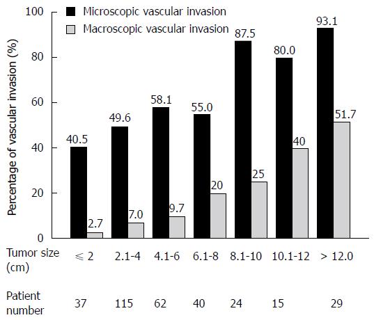
Figure 3 Frequency of microscopic and macroscopic vascular invasion in 322 patients with hepatocellular carcinoma stratified according to tumor size.
(From Tsai et al[76]).
- Citation: Chau GY. Resection of hepatitis B virus-related hepatocellular carcinoma: Evolving strategies and emerging therapies to improve outcome. World J Gastroenterol 2014; 20(35): 12473-12484
- URL: https://www.wjgnet.com/1007-9327/full/v20/i35/12473.htm
- DOI: https://dx.doi.org/10.3748/wjg.v20.i35.12473


