Copyright
©2014 Baishideng Publishing Group Inc.
World J Gastroenterol. Aug 28, 2014; 20(32): 11216-11229
Published online Aug 28, 2014. doi: 10.3748/wjg.v20.i32.11216
Published online Aug 28, 2014. doi: 10.3748/wjg.v20.i32.11216
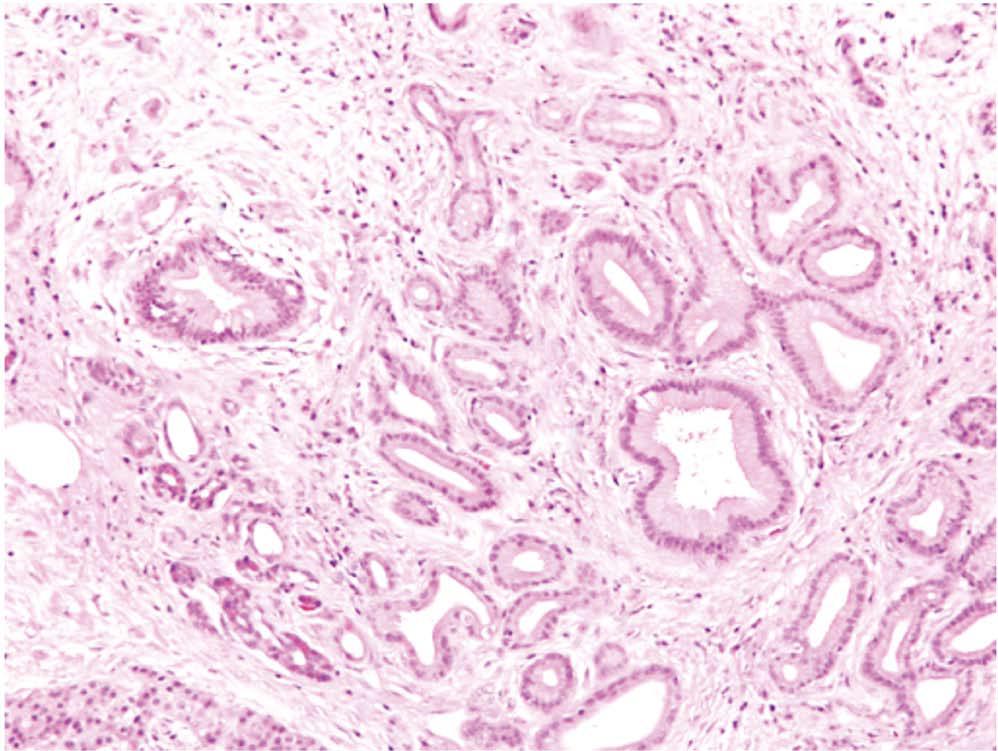
Figure 1 Pancreatic cancer and stromal reaction.
A representative HE stained human pancreatic cancer tissue section showing duct-like and tubular structures (malignant elements, examples highlighted by arrows) infiltrating into and embedded in a highly fibrotic stromal reaction (examples highlighted by asterisks).
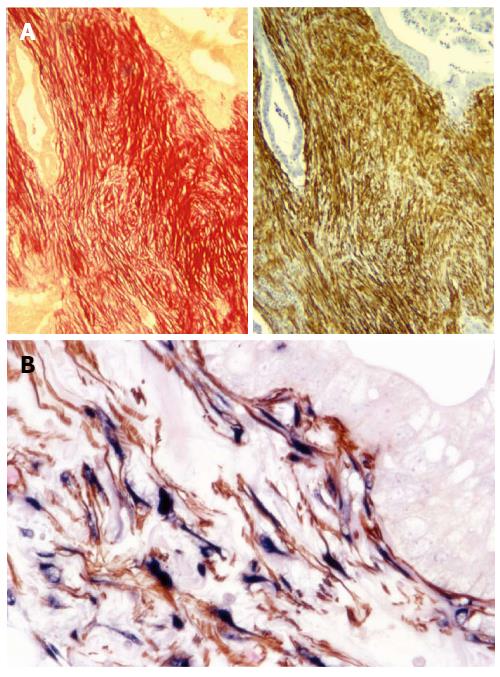
Figure 2 Pancreatic stellate cells are the source of collagen in stroma.
A: A representative pair of serial sections of human pancreatic cancer tissue shows that Sirius Red staining for collagen (red) co-localises with immunohistochemical staining for α-smooth muscle actin (α-SMA) (brown), suggesting the presence of activated pancreatic stellate cells (PSCs) in the stroma of pancreatic cancer[33]. Reprinted with permission from Wolters Kluwer Health (Apte et al[33]); B: Immunohistochemistry for α-SMA (brown) and in situ hybridisation for procollagen α1 mRNA (blue), reveals colocalisation of α-SMA and procollagen mRNA on human pancreatic cancer tissue indicating that active PSCs are the major source of collagen in tumour stroma. Reprinted with permission from Elsevier (Apte et al[43]).
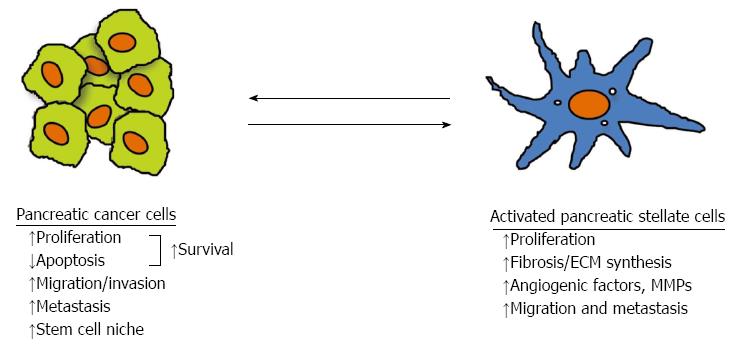
Figure 3 Interaction between pancreatic cancer cells and pancreatic stellate cells.
Pancreatic cancer cells stimulate the proliferation, extracellular matrix (ECM) production, angiogenic factors and matrix metalloproteinase (MMP) expression, as well as migration of pancreatic stellate cells (PSCs); conversely PSCs increase proliferation and reduce apoptosis leading to increased survival, increase cancer cell migration and facilitate a cancer stem cell niche. The overall effect of interaction between pancreatic cancer cells and PSCs facilitates cancer progression[43].
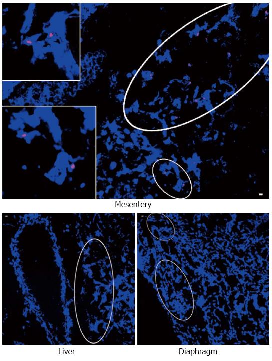
Figure 4 Identification of human pancreatic stellate cells from primary tumour in metastatic nodules.
A representative photomicrograph showing Y chromosome positive cells in metastatic nodules in the mesentery (inserts are high power views of the circled regions), liver and diaphragm from mice (female) injected with female pancreatic cancer cells + male human pancreatic stellate cells, using fluorescent in situ hybridisation for the Y chromosome. Reprinted with permission from Elsevier (Xu et al[63]).
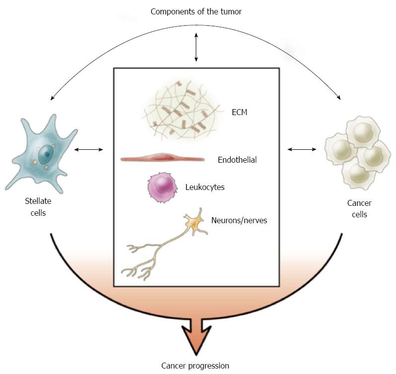
Figure 5 Tumour components.
The stromal reaction of pancreatic ductal adenocarcinoma is comprised of pancreatic stellate cells (stromal cells), abundant extracellular matrix (ECM), blood vessels/endothelial cells, immune cells and nerves/neurons[43]. The interaction between cancer cells and the components of stroma facilitates cancer progression. Reprinted with permission from Elsevier (Apte et al[43]).
- Citation: Xu Z, Pothula SP, Wilson JS, Apte MV. Pancreatic cancer and its stroma: A conspiracy theory. World J Gastroenterol 2014; 20(32): 11216-11229
- URL: https://www.wjgnet.com/1007-9327/full/v20/i32/11216.htm
- DOI: https://dx.doi.org/10.3748/wjg.v20.i32.11216


