Copyright
©2014 Baishideng Publishing Group Inc.
World J Gastroenterol. Aug 28, 2014; 20(32): 11095-11115
Published online Aug 28, 2014. doi: 10.3748/wjg.v20.i32.11095
Published online Aug 28, 2014. doi: 10.3748/wjg.v20.i32.11095
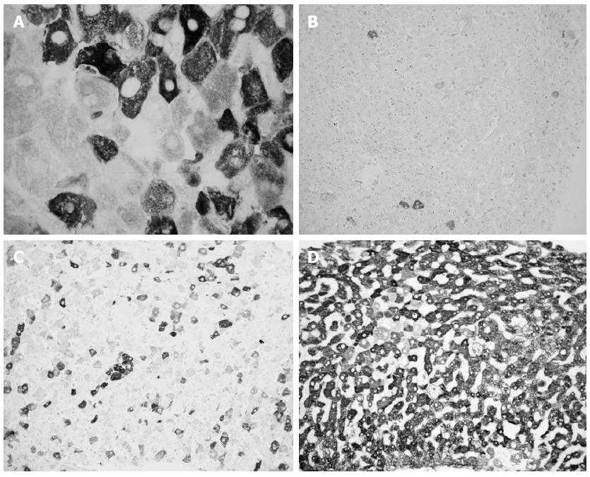
Figure 1 Immunohistochemistry of lobular areas from different liver biopsies stained for hepatitis C virus-antigens.
Cytoplasmic positivity of hepatocytes with different intensities of staining. A: Negative and strongly positive hepatocytes in the same areas. Original magnification 120 ×; B: Few positive hepatocytes. Original magnification 20 ×; C: About 20% positive hepatocytes with different intensity of staining. Original magnification 20 ×; D: Widespread positivity, with prevalent strong intensity of staining. Original magnification 20 ×.
- Citation: Grassi A, Ballardini G. Post-liver transplant hepatitis C virus recurrence: An unresolved thorny problem. World J Gastroenterol 2014; 20(32): 11095-11115
- URL: https://www.wjgnet.com/1007-9327/full/v20/i32/11095.htm
- DOI: https://dx.doi.org/10.3748/wjg.v20.i32.11095


