Copyright
©2014 Baishideng Publishing Group Inc.
World J Gastroenterol. Aug 21, 2014; 20(31): 10864-10875
Published online Aug 21, 2014. doi: 10.3748/wjg.v20.i31.10864
Published online Aug 21, 2014. doi: 10.3748/wjg.v20.i31.10864
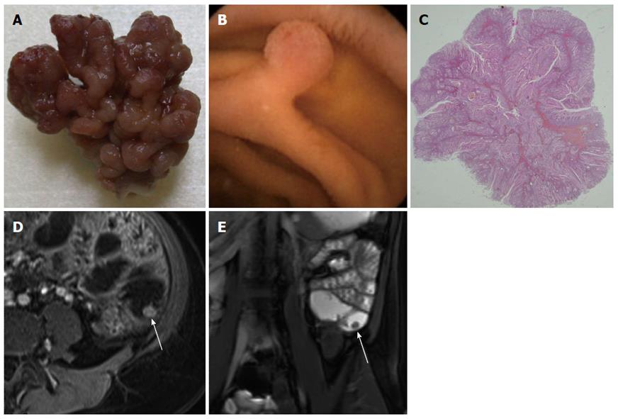
Figure 1 Polyp 1 in a 46-year-old female with known peutz-Jeghers syndrome.
A: Macroscopically, the peutz-Jeghers syndrome (PJS) polyp has a coarse lobulated surface; B: The capsule endoscopy image shows that this polyp is pediculate; C: Microscopically, the analysis confirms a benign hamartomatous polyp with a tree-like branching core derived from the muscularis mucosae covered by normal epithelium; D: Fat-suppressed axial T1-weighted-Gadolinium-enhanced VIBE image shows a moderate enhancement of the polyp, with a good characterization of its size and shape (arrow); E: The coronal T2 True Fast Imaging with Steady-state in Precession (FISP) MR image also allows detection of this isolated ileal polyp (arrow).
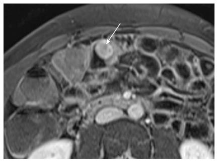
Figure 2 Polyp 2 in a 26-year-old female with known peutz-Jeghers syndrome.
Axial fat-suppressed T1 weighted-Gadolinium-enhanced axial image shows the enhancement of the rounded PJS polyp (arrow).
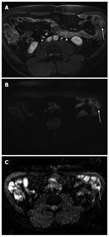
Figure 3 Polyp 3 in a 54-year-old male with known peutz-Jeghers syndrome.
A: Axial fat-saturated Vibe gadolinium-enhanced T1-weighted shows a small-bowel slightly enhancing jejunal polyp (arrow); B, C: The corresponding images on a high b (b = 800) value diffusion-weighted MRI shows a high-signal intensity polypoid lesion inside a jejunal small-bowel loop with a low ADC value compared with the surrounding small-bowel lumen (ADC = 1440 mm2/s) (arrow).
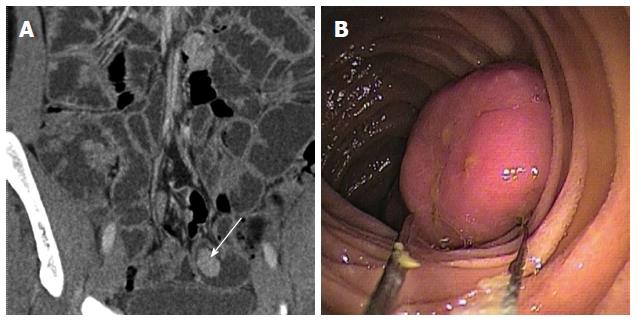
Figure 4 Polyp 4.
A: Multidetector computed tomography (MDCT) coronal view reveals a regular small-bowel polyp with homogeneous enhancement (arrow); B: The double balloon endoscopy optimally depicts this large small bowel polyp.
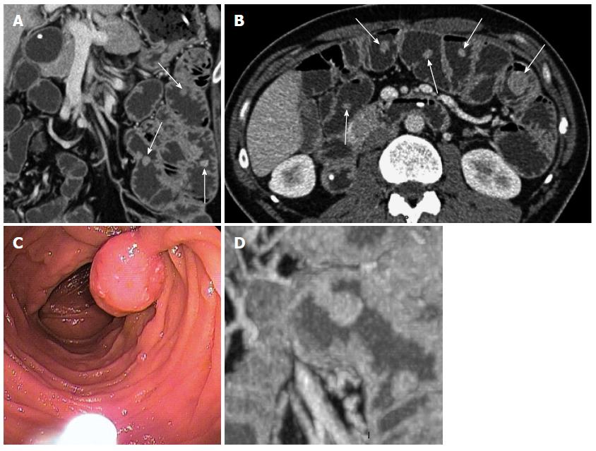
Figure 5 Polyp 5 in a 44-year-old male with a known peutz-Jeghers syndrome.
A: Coronal and B: an axial Multidetector computed tomography (MDCT) enteroclysis images reveals multiple, regular polyps with various sizes and shapes (arrows); C: The endoscopic view shows a pediculate polyp; D: A coronal MIP reformat shows the typical pediculate peutz-Jeghers syndrome (PJS) polyp aspect.
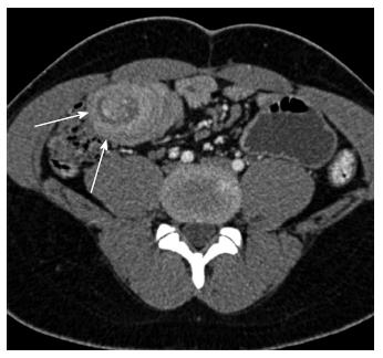
Figure 6 Twenty-four-year-old male referred at the emergency department for abdominal pain.
The axial Multidetector computed tomography (MDCT) scan shows the bowel-in-bowel appearance with mesenteric fat into the intussusception (arrows).
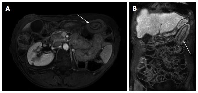
Figure 7 Axial (A) and (B) coronal fat-saturated Vibe gadolinium-enhanced T1-weighted magnetic resonance images illustrate the target-like or sausage shape of the small-bowel intussusception.
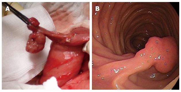
Figure 8 Operative view shows a pediculate polyp that was removed (A) (reprinted with permission from ref.
38) after double balloon enteroscopic evaluation (B).
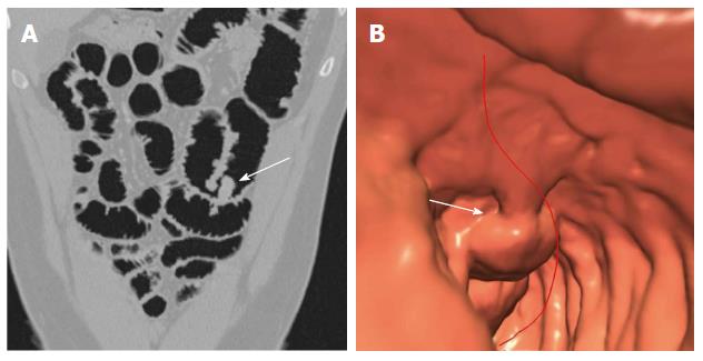
Figure 9 Twenty-eight-year-old male with a known peutz-Jeghers syndrome presenting a single small-bowel polyp.
A: Multidetector row computed tomography-virtual endoscopy (MDCT-VE) acquired after gas filling shows a single polyp in the intestinal lumen (arrow); B: Corresponding image of this polyp after MDCT-VE post-processing (arrow).
- Citation: Tomas C, Soyer P, Dohan A, Dray X, Boudiaf M, Hoeffel C. Update on imaging of Peutz-Jeghers syndrome. World J Gastroenterol 2014; 20(31): 10864-10875
- URL: https://www.wjgnet.com/1007-9327/full/v20/i31/10864.htm
- DOI: https://dx.doi.org/10.3748/wjg.v20.i31.10864


