Copyright
©2014 Baishideng Publishing Group Inc.
World J Gastroenterol. Aug 21, 2014; 20(31): 10864-10875
Published online Aug 21, 2014. doi: 10.3748/wjg.v20.i31.10864
Published online Aug 21, 2014. doi: 10.3748/wjg.v20.i31.10864
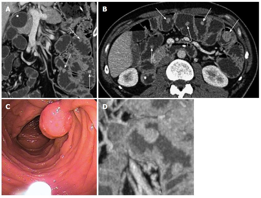
Figure 5 Polyp 5 in a 44-year-old male with a known peutz-Jeghers syndrome.
A: Coronal and B: an axial Multidetector computed tomography (MDCT) enteroclysis images reveals multiple, regular polyps with various sizes and shapes (arrows); C: The endoscopic view shows a pediculate polyp; D: A coronal MIP reformat shows the typical pediculate peutz-Jeghers syndrome (PJS) polyp aspect.
- Citation: Tomas C, Soyer P, Dohan A, Dray X, Boudiaf M, Hoeffel C. Update on imaging of Peutz-Jeghers syndrome. World J Gastroenterol 2014; 20(31): 10864-10875
- URL: https://www.wjgnet.com/1007-9327/full/v20/i31/10864.htm
- DOI: https://dx.doi.org/10.3748/wjg.v20.i31.10864


